. 2010 Oct;30(20):4818-27.
doi: 10.1128/MCB.01348-09. Epub 2010 Aug 16.

The transcriptional mediator subunit MED1/TRAP220 in stromal cells is involved in hematopoietic stem/progenitor cell support through osteopontin expression

Affiliations

The transcriptional mediator subunit MED1/TRAP220 in stromal cells is involved in hematopoietic stem/progenitor cell support through osteopontin expression

Akiko Sumitomo et al. Mol Cell Biol. 2010 Oct.

Abstract

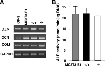
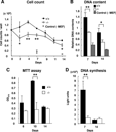
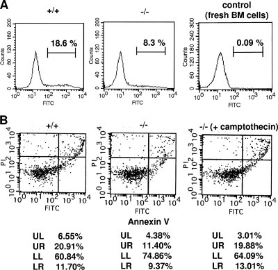
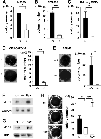
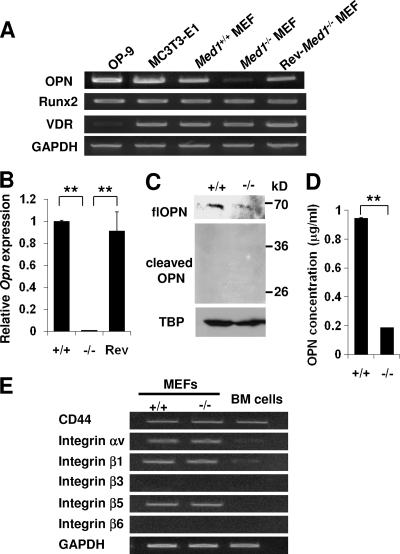
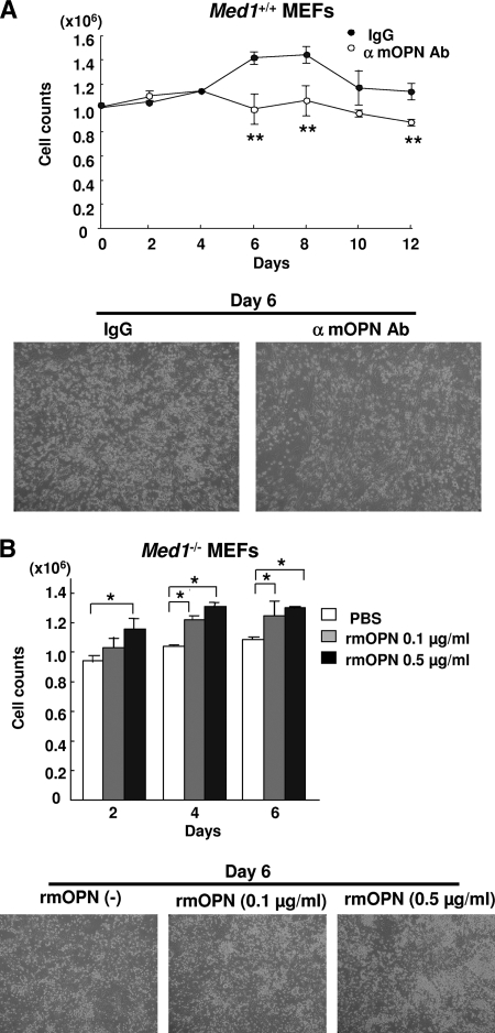
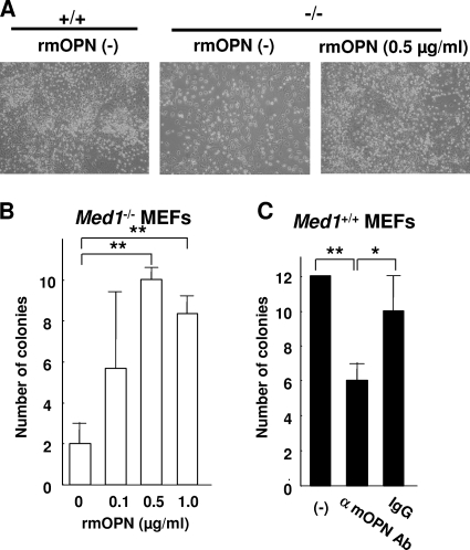
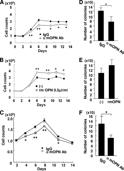
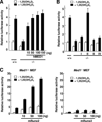
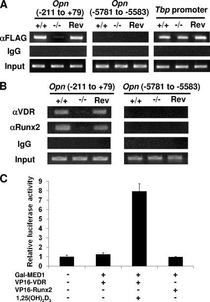
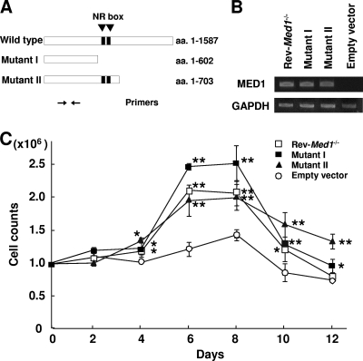
MED1/TRAP220, a subunit of the transcriptional Mediator/TRAP complex, is crucial for various biological events through its interaction with distinct activators, such as nuclear receptors and GATA family activators. In hematopoiesis, MED1 plays a pivotal role in optimal nuclear receptor-mediated myelomonopoiesis and GATA-1-induced erythropoiesis. In this study, we present evidence that MED1 in stromal cells is involved in supporting hematopoietic stem and/or progenitor cells (HSPCs) through osteopontin (OPN) expression. We found that the proliferation of bone marrow (BM) cells cocultured with MED1 knockout (Med1(-/-)) mouse embryonic fibroblasts (MEFs) was significantly suppressed compared to the control. Furthermore, the number of long-term culture-initiating cells (LTC-ICs) was attenuated for BM cells cocultured with Med1(-/-) MEFs. The vitamin D receptor (VDR)- and Runx2-mediated expression of OPN, as well as Mediator recruitment to the Opn promoter, was specifically attenuated in the Med1(-/-) MEFs. Addition of OPN to these MEFs restored the growth of cocultured BM cells and the number of LTC-ICs, both of which were attenuated by the addition of the anti-OPN antibody to Med1(+/+) MEFs and to BM stromal cells. Consequently, MED1 in niche appears to play an important role in supporting HSPCs by upregulating VDR- and Runx2-mediated transcription on the Opn promoter.


Figures

FIG. 1.
FIG. 1.
MEFs express molecules attributable to the osteoblastic lineage. (A) Both Med1+/+ and Med1−/− MEFs (p53−/−), as well as MC3T3-E1 and OP-9 cells, express mRNAs of ALP, osteocalcin (OCN), and COLI. (B) The ALP activities of Med1+/+ and Med1−/− MEF lysates are comparable to those of MC3T3-E1 cells. The values are means ± standard deviations (SD) of a representative experiment performed in duplicate.
FIG. 2.
FIG. 2.
MED1 in MEFs mediates mitogenic stress to cocultured BM cells. (A and B) The numbers (A) and DNA content (B) of the total BM cells cultured on the Med1−/− MEFs are less than those of cells cultured on Med1+/+ MEFs but more than those of cells cultured without MEFs. (C and D) The number of live BM cells, measured by the MTT assay (C), and the level of BM cell DNA synthesis, measured by BrdU incorporation (D), were less when cells were cocultured on Med1−/− MEFs than when they were cultured on Med1+/+ MEFs. The values are means ± SD of a representative experiment performed in triplicate (*, P < 0.05; **, P < 0.01).
FIG. 3.
FIG. 3.
BM cells on Med1−/− MEFs are less susceptible to cell death. (A) Incorporation of FITC-dUTP into BM cells by TdT. Normal BM cells were used as the negative control. (B) Annexin V-FITC and PI double staining. Cells treated with 50 nM camptothecin for 4 h were used as the positive control. BM cells cocultured for a week on Med1+/+ MEFs are more susceptible to apoptosis (A and B; LR) and necrosis (B; UR).
FIG. 4.
FIG. 4.
MED1 in MEFs mediates support of LTC-ICs. (A and B) After a 6-week coculture, the numbers of CFCs on Med1+/+ MEFs (p53−/−) in M5300 medium (A) and BIT9500-based medium (B) are higher than the numbers of CFCs on Med1−/− MEFs (p53−/−). (C) After a 6-week coculture, the number of CFCs on primary Med1+/+ MEFs (p53+/+) is higher than numbers of CFCs on primary Med1−/− MEFs (p53+/+). (D and E) The numbers of myeloid (D) and erythroid (E) colonies on Med1+/+ MEFs (p53−/−) in M5300 medium are higher than the numbers of colonies on Med1−/− MEFs (p53−/−). (F and G) Northern (F) and Western (G) blot analyses of the Med1+/+, Med1−/−, and Rev-Med1−/− MEFs. GAPDH (F) and TATA-binding protein (TBP) (G) were used as a control. (H) The CFCs on the Rev-Med1−/− MEFs recovered to the level on the Med1+/+ MEFs. The values are means ± SD of a representative experiment performed in triplicate (*, P < 0.05; **, P < 0.01).
FIG. 5.
FIG. 5.
Expression of flOPN is attenuated in Med1−/− MEFs. (A and B) Semiquantitative (A) and quantitative (B) PCR. Opn mRNA is suppressed in the Med1−/− MEFs but recovers in the Rev-Med1−/− MEFs. (C) Western blot analysis. Expression of flOPN is reduced in the Med1−/− MEFs; trOPN is invisible. (D) ELISA of OPN. OPN concentrations in culture media during LTC-IC assays were measured. OPN produced by the Med1−/− MEFs is reduced. (E) Expression of various receptors for OPN in MEFs and BM cells was analyzed by semiquantitative PCR.
FIG. 6.
FIG. 6.
OPN mediates mitogenicity of BM cells on MEFs. (A) The number of BM cells cocultured on Med1+/+ MEFs in the presence of the anti-mOPN antibody (Ab) is attenuated. α, anti. (B) The number of BM cells cocultured on Med1−/− MEFs increases in an rmOPN dose-dependent manner. PBS, phosphate-buffered saline. The values are the means ± SD of a representative experiment performed in triplicate (*, P < 0.05; **, P < 0.01).
FIG. 7.
FIG. 7.
OPN mediates the support of LTC-ICs on MEFs. (A) The number of BM cells on Med1−/− MEFs in the presence of rmOPN recovers to the control level. (B) The number of CFCs on Med1−/− MEFs increases in the presence of rmOPN in a dose-dependent manner. (C) The number of CFCs on Med1+/+ MEFs is reduced in the presence of the anti-mOPN antibody. The values are the means ± SD of a representative experiment performed in triplicate (*, P < 0.05; **, P < 0.01).
FIG. 8.
FIG. 8.
OPN depletion attenuates the mitogenicity of BM cells and LTC-ICs cocultured on BM stromal cells. (A to C) BM cells on MS-5 (A and B) and OP-9 (C) BM stromal cells during a 2-week period were counted in the presence of the anti-mOPN antibody (A and C) or rmOPN (B). (D to F) CFCs after a 6-week coculture on MS-5 (D and E) and OP-9 (F) BM stromal cells were counted in the presence of the anti-mOPN antibody (D and F) or rmOPN (E). The numbers of BM cells and CFCs on stromal cells are reduced in the presence of the anti-mOPN antibody, and the number of BM cells increases in the presence of rmOPN. The values are the means ± SD of a representative experiment performed in triplicate (*, P < 0.05; **, P < 0.01).
FIG. 9.
FIG. 9.
MED1 mediates VDR- and Runx2-mediated transcription on the Opn promoter. Luciferase reporter assays were performed in the presence of VDR. (A) Ligand-dependent activation is attenuated in the Med1−/− MEFs but recovers in a MED1 dose-dependent manner. (B) Reintroduction of mutants I [MED1(1-602)]) and IV (MED1 with NR box mutations) into Med1−/− MEFs recovers the basal level of transcription but not ligand-dependent activation, whereas reintroduction of mutant II [MED1(1-703)] recovers both basal transcription and ligand-induced activation. Mutant III [MED(592-1587)] has no effect. (C) Addition of Runx2 activates both ligand-independent and -dependent activation in the Med1+/+ MEFs but not in the Med1−/− MEFs. The values (means ± SD of a representative experiment performed in triplicate) are plotted as a fold increases against the value for Med1+/+ MEFs without a ligand.
FIG. 10.
FIG. 10.
Recruitment of MED1, VDR, and Runx2 on the Opn promoter. (A and B) ChIP assays. (A) FLAG-MED10 was transiently expressed, and sheared chromatin was immunoprecipitated with anti-FLAG (M2) IgG or mouse IgG as the control. On the Opn promoter proximal to the transcription initiation point, Mediator recruitment is attenuated in the Med1−/− MEFs but recovered in the Rev-Med1−/− MEFs. Mediator is not recruited to the 5′ remote region. Mediator recruitment on the Tbp promoter is comparable. (B) VDR and Runx2 recruitment is attenuated in the Med1−/− MEFs but recovers in the Rev-Med1−/− MEFs. (C) Mammalian two-hybrid assays. Luciferase activities of the reporter with 5 Gal4-binding sites were measured. Gal4-fused MED1 interacts with VP16-fused VDR in a ligand-dependent manner, but not with VP16-fused Runx2.
FIG. 11.
FIG. 11.
MED1(1-602) in MEFs is sufficient for the mitogenicity of BM cells. (A) Primer positions for PCR. (B) Semiquantitative PCR shows stable expression of the MED1 mutants in the Med1−/− MEFs. (C) Mutants I and II as well as the Rev-Med1−/− MEFs recover the mitogenicity of BM cells. The values are the means ± standard errors (SE) of a representative experiment performed in triplicate (*, P < 0.05; **, P < 0.01).

Similar articles

Cited by

References

    1. Adams, G. B., and D. T. Scadden. 2006. The hematopoietic stem cell in its place. Nat. Immunol. 7:333-337. - PubMed
    1. Askmyr, M., N. A. Sims, T. J. Martin, and L. E. Purton. 2009. What is the true nature of the osteoblastic hematopoietic stem cell niche? Trends Endocrinol. Metab. 20:303-309. - PubMed
    1. Chang, P. L., M. Cao, and P. Hicks. 2003. Osteopontin induction is required for tumor promoter-induced transformation of preneoplastic mouse cells. Carcinogenesis 24:1749-1758. - PubMed
    1. Conaway, R. C., S. Sato, C. Tomomori-Sato, T. Yao, and J. W. Conaway. 2005. The mammalian mediator complex and its role in transcriptional regulation. Trends Biochem. Sci. 30:250-255. - PubMed
    1. Fan, X., D. M. Chou, and K. Struhl. 2006. Activator-specific recruitment of Mediator in vivo. Nat. Struct. Mol. Biol. 13:117-120. - PubMed

Publication types

MeSH terms