. 2010 Oct;30(20):4952-64.
doi: 10.1128/MCB.00299-10. Epub 2010 Aug 16.

Regulation of nucleolar chromatin by B23/nucleophosmin jointly depends upon its RNA binding activity and transcription factor UBF

Affiliations

Regulation of nucleolar chromatin by B23/nucleophosmin jointly depends upon its RNA binding activity and transcription factor UBF

Miharu Hisaoka et al. Mol Cell Biol. 2010 Oct.

Abstract

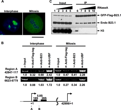
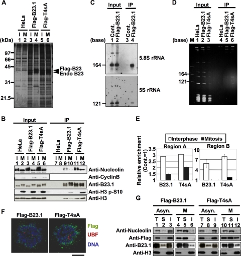
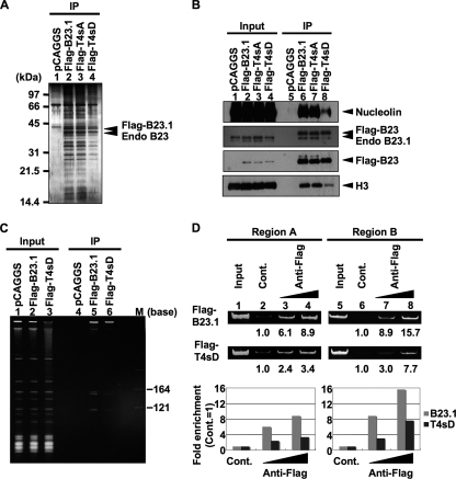
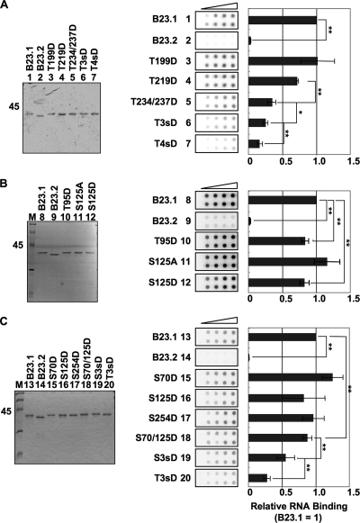
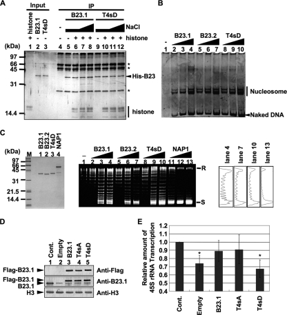
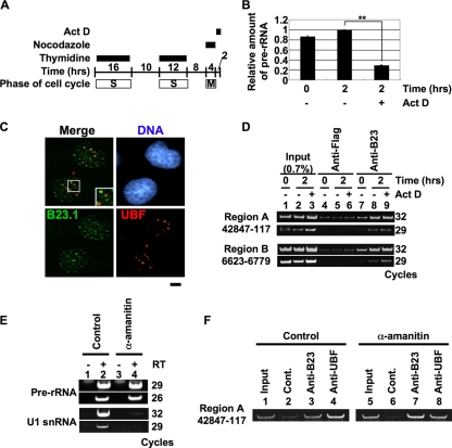
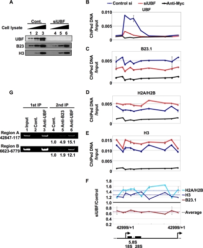
Histone chaperones regulate the density of incorporated histone proteins around DNA transcription sites and therefore constitute an important site-specific regulatory mechanism for the control of gene expression. At present, the targeting mechanism conferring this site specificity is unknown. We previously reported that the histone chaperone B23/nucleophosmin associates with rRNA chromatin (r-chromatin) to stimulate rRNA transcription. Here, we report on the mechanism for site-specific targeting of B23 to the r-chromatin. We observed that, during mitosis, B23 was released from chromatin upon inactivation of its RNA binding activity by cdc2 kinase-mediated phosphorylation. The phosphorylation status of B23 was also shown to strongly affect its chromatin binding activity. We further found that r-chromatin binding of B23 was a necessary condition for B23 histone chaperone activity in vivo. In addition, we found that depletion of upstream binding factor (UBF; an rRNA transcription factor) decreased the chromatin binding affinity of B23, which in turn led to an increase in histone density at the r-chromatin. These two major strands of evidence suggest a novel cell cycle-dependent mechanism for the site-specific regulation of histone density via joint RNA- and transcription factor-mediated recruitment of histone chaperones to specific chromosome loci.


Figures

FIG. 1.
FIG. 1.
B23.1 is released from chromatin during mitosis. (A) Localization of GFP-Flag-B23.1. HeLa cells stably expressing GFP-Flag-B23.1 grown on coverslips were fixed with 3% paraformaldehyde, and the localization of the protein was observed under a confocal microscope. DNA was counterstained with TO-PRO-3. Bar, 10 μm. (B) ChIP assays of endogenous B23. Asynchronous and mitotic HeLa cells were subjected to ChIP assays using anti-Flag-tag as a control, anti-B23, and anti-UBF antibodies. The input (lanes 1 and 5) and coimmunoprecipitated (lanes 2 to 4 and 6 to 8) DNA were purified, and the DNA fragment harboring the rRNA gene promoter (region A; nucleotide positions between 42847 (−153) and 117 relative to the transcription start site [+1]) and coding (region B; nucleotide positions 6623 to 6779) regions were amplified by PCR. PCR products were analyzed by 6% PAGE and visualized with GelRed staining. The amounts of PCR products amplified from input DNA were set as 1.0, and the enrichment is shown at the bottom of the panel. The positions of primers on the rRNA gene are schematically represented at the bottom. (C) Immunoprecipitation of GFP-Flag-B23.1 with RNase-treated cell extracts. HeLa cells expressing GFP-Flag-B23.1 were treated without (lanes 1 and 6) or with 1, 10, 100, or 1,000 μg/ml of RNase A (lanes 2 to 5 and 7 to 10). GFP-Flag-B23.1 was immunoprecipitated with anti-Flag-tag antibody and separated by SDS-PAGE followed by Western blotting with anti-B23.1 and anti-histone H3 antibodies.
FIG. 2.
FIG. 2.
Mitotic phosphorylation of B23.1 is required for the inactivation of its chromatin and RNA binding activities. (A and B) Immunoprecipitation of Flag-tagged proteins. Cell extracts were prepared from asynchronous (I) or mitotic (M) HeLa cells and HeLa cells expressing either Flag-B23.1 or -T4sA, and immunoprecipitation using anti-Flag-tag antibody was carried out. Immunoprecipitated proteins were separated by SDS-PAGE and detected by silver staining (A) and Western blotting (B). Input and immunoprecipitated proteins (A, lanes 1 to 6 and 7 to 12, respectively) were analyzed by Western blotting using anti-B23, antinucleolin, anti-histone H3 S10P (histone H3 phosphorylated at serine 10), and anti-histone H3 antibodies. Western blotting with anti-cyclin B antibody was used as a mitotic marker (B, second panel). (C) RNAs coprecipitated with Flag-B23.1. Nuclear extracts were prepared from control and Flag-B23.1-expressing HeLa cells, and immunoprecipitation with anti-Flag-tag antibody was carried out. The input and coimmunoprecipitated RNA was purified, separated on a 6% denaturing PAGE gel, and analyzed by Northern blotting with 5S and 5.8S rRNA gene probes. (D) RNA binding activity of B23.1 and T4sA during mitosis. Cell extracts were prepared from mitotic HeLa cells expressing either Flag-B23.1 or -T4sA, as described in Materials and Methods, and immunoprecipitation with anti-Flag-tag antibody was carried out. Coimmunoprecipitated RNAs were purified, separated on a 6% denaturing PAGE gel, and detected with GelRed staining. Lane M indicates RNA molecular markers. (E) Cell cycle-dependent association of B23.1 and T4sA with the rRNA gene. Asynchronous and mitotic HeLa cells expressing either Flag-B23.1 or -T4sA were subjected to ChIP assays using control IgG and anti-Flag-tag antibody. Coimmunoprecipitated and input DNA were used as templates for Q-PCR using the primer sets for both regions A and B. The amount of amplified DNA precipitated with control IgG was set as 1.0, and the relative enrichment level of the DNA fragments by anti-Flag-tag immunoprecipitation are shown. White and black bars in the graph show the results of ChIP assays with asynchronous and mitotic cells, respectively. Q-PCRs were carried out in triplicate. (F) Localization of B23.1 and T4sA in mitotic cells. HeLa cells expressing either Flag-B23.1 or -T4sA grown on coverslips (left and right panels, respectively) were fixed in 1% paraformaldehyde, and Flag-tagged proteins (green) and UBF (red) were detected by specific antibodies. DNA was counterstained with TO-PRO-3 (blue). Localization of proteins was observed under a confocal microscope. Bar, 10 μm. (G) Extractability of B23.1 and T4sA in asynchronous and mitotic cells. Asynchronous or mitotic (lanes 1 to 3 and 7 to 9 or 4 to 6 and 10 to 12, respectively) HeLa cells expressing either Flag-B23.1 or -T4sA were fractionated to soluble and insoluble fractions. Total cell extracts and soluble and insoluble fractions (indicated by T, S, and I, respectively) were separated by SDS-PAGE and analyzed by Western blotting using antibodies against nucleolin, Flag tag, B23.1, and histone H3.
FIG. 3.
FIG. 3.
The phosphomimetic B23.1 mutant does not efficiently associate with chromatin. (A and B) Immunoprecipitation of Flag-tagged B23.1 proteins. 293T cells were transfected with empty vector or vectors for expression of Flag-B23.1, -T4sA, and -T4sD (lanes 1 to 4, respectively), and immunoprecipitation was carried out with anti-Flag-tag antibody. Precipitated proteins were separated by SDS-PAGE and detected with silver staining (A) or Western blotting (B). Western blotting was carried out with anti-Flag-tag, -nucleolin, -B23, and -histone H3 antibodies. (C) RNA binding activity of T4sD in 293T cells. Cell extracts were prepared as described in Materials and Methods. RNAs coimmunoprecipitated with Flag-tagged proteins were separated on a 6% denaturing PAGE gel and visualized with staining by GelRed. Lane M indicates RNA molecular markers. (D) r-chromatin association of B23.1 and T4sD. Flag-B23.1 or -T4sD was transiently expressed in 293T cells, and ChIP assays using anti-Flag-tag antibody (0.5 and 1.0 μg) were carried out. Anti-Myc-tag antibody (1 μg) was used as a control. Precipitated and input DNA were used as templates for PCR with specific primer sets (regions A and B). PCR product from control immunoprecipitation was set as 1.0, and the relative enrichment levels of DNA fragments by anti-Flag-tag immunoprecipitation are indicated at the bottom and graphically represented. Gray and black bars in the graphs show the results of ChIP assays with Flag-B23.1 and -T4sD, respectively.
FIG. 4.
FIG. 4.
Phosphorylation of four cdc2 consensus sites of B23 is crucial for efficient suppression of its RNA binding activity. (A) RNA binding activity of B23 proteins containing mutations at cdc2 consensus sites. B23.1, B23.2, T199D, T219D, T234/237D, T3sD, and T4sD proteins (lanes 1 to 7, respectively) (200 ng) were separated by SDS-PAGE and visualized by Coomassie brilliant blue (CBB) staining (left). T3sD and T4sD are B23.1 mutants in which T219, T234, and T237 and all four threonines (T), respectively, are replaced with aspartic acids (D). Purified proteins (50, 100, 200, and 400 ng) were mixed with 32P-labeled HeLa cell total RNA (10 ng). The mixture was incubated and then filtered though nitrocellulose membranes. The membranes were extensively washed, and RNA retained on the membrane was detected with an image analyzer (middle). The intensity of each spot was analyzed, and the RNA binding activity obtained with the same amount of B23 proteins was first calculated relative to that of B23.1 (1.0). Then the relative RNA binding activity at each protein amount was averaged. Means ± standard deviations (SD) obtained from two duplicate independent experiments are shown (center and right). (B) The effects of T95 and S125 phosphorylation on the RNA binding activity of B23.1. Purified proteins B23.1, B23.2, T95D, S125A, and S125D (200 ng) were separated by SDS-PAGE (lanes 8 to 12, respectively) and visualized with CBB staining. The RNA binding activity of B23 mutant proteins was examined by filter binding assays as described for panel A. (C) The effects of the position and number of phosphorylation sites on the RNA binding activity of B23.1. Purified proteins B23.1, B23.2, S70D, S125D, S254D, S70/125D, S3sD (S70/125/254D), and T3sD (T219/234/237D) (200 ng) were separated by SDS-PAGE (lanes 13 to 20, respectively) and visualized with CBB staining. The RNA binding activity of each protein was examined as described for panel A. For all experiments, statistical P values were calculated by t tests and indicated with * for P < 0.05 and ** for P < 0.01.
FIG. 5.
FIG. 5.
RNA binding activity of B23.1 is required to facilitate rRNA transcription. (A) Histone binding activity of recombinant proteins. Recombinant B23.1 or T4sD (1 μg) was preincubated without (lanes 5 and 9) or with core histones (300 ng, lanes 6 to 8 and 10 to 12), and immunoprecipitation was carried out with anti-B23.1 antibody. Bound proteins were washed in a buffer containing 150 mM (lanes 4 to 6 and 9 to 10), 250 mM (lanes 7 and 11), or 400 mM (lanes 8 and 12) NaCl and were separated by SDS-PAGE followed by silver staining. The asterisks indicate bands derived from the antibody. (B) The histone transfer activity of B23 proteins. Core histones (72 ng) were preincubated without (lane 1) or with 50 (lanes 2, 5, and 8), 150 (lanes 3, 6, and 9), or 450 (lanes 4, 7, and 10) ng of recombinant B23.1, B23.2, or T4sD (lanes 2 to 4, 5 to 7, and 8 to 10, respectively) and then mixed with the 147-bp-long DNA fragment and further incubated. The mixtures were separated on a 6% PAGE gel, and DNA was visualized with GelRed staining. (C) Supercoiling assays. B23.1, B23.2, T4sD, and NAP1 proteins were separated by SDS-PAGE and visualized by CBB staining (left). Core histones (1.8 pmol) were preincubated without (lane 1) or with 1.8 (lanes 2, 5, 8, and 11), 5.4 (lanes 3, 6, 9, and 12), or 16.2 (lanes 4, 7, 10, and 13) pmol of recombinant B23.1, B23.2, T4sD, or NAP1 (lanes 2 to 4, 5 to 7, 8 to 10, or 11 to 13, respectively) and then mixed with plasmid DNA preincubated with topoisomerase I and further incubated. DNA was purified and separated on a 1% agarose gel and visualized with GelRed staining. Positions of relaxed (R) or supercoiled (S) circular plasmid DNA are indicated. The band intensities of lanes 4, 7, 10, and 13 were quantified and plotted (right). (D) Expression level of endogenous and exogenous B23 proteins. HeLa cells were transfected with control or B23.1 siRNAs. The cells were supertransfected 24 h after siRNA transfection with empty vector or vectors expressing either Flag-B23.1, -T4sA, or -T4sD. An equal number of cells was collected 72 h after siRNA transfection and subjected to Western blotting with anti-Flag-tag, -B23.1, and -histone H3 antibodies. (E) rRNA transcription level of siRNA-treated cells. Total RNA was isolated from 7 × 10⁵ cells prepared as described for panel D, and the rRNA transcription level was examined by quantitative RT-PCR using a 5′-ETS-specific primer set. The amount of pre-rRNA was normalized by that of β-actin mRNA. Results are means ± SD obtained from three independent experiments. Statistical P values were calculated by t tests and are indicated with * for P < 0.05.
FIG. 6.
FIG. 6.
Nascent rRNA is not crucial for the recruitment of the B23-RNA complex to r-chromatin. (A to C) Effect of Act D on rRNA transcription. HeLa cells were synchronized at mitosis and released for 2 h in growth medium in the absence or presence of 50 ng/ml Act D (A). Total RNAs were purified, and the rRNA transcription level was examined by quantitative RT-PCR using a 5′-ETS-specific primer set (B). The amount of pre-rRNA was normalized by that of β-actin mRNA. Results are means ± SD obtained from three independent experiments. Statistical P values are indicated with ** for P < 0.01. HeLa cells prepared as described for panel A were fixed and subjected to immunofluorescence analysis with anti-B23.1 and anti-UBF antibodies (C). DNA was counterstained with TO-PRO-3. Bar, 5 μm. (D) Effect of rRNA transcription initiation on the recruitment of B23 to r-chromatin. HeLa cells prepared as described for panel A were subjected to ChIP assays using anti-Flag-tag and -B23 (lanes 4 to 6 and 7 to 9, respectively) antibodies. Precipitated (lanes 4 to 9) and input (lanes 1 to 3) DNA were used for PCR with region A and B primer sets. PCR products were analyzed on a 6% PAGE gel and visualized with GelRed staining. (E) Effect of α-amanitin on transcription. HeLa cells were cultured in the absence or presence of 5 μg/ml of α-amanitin for 24 h, and total RNA was isolated from 4.5 × 10⁵ cells. The amounts of pre-rRNA and U1 snRNA were examined by RT-PCR. PCR products were analyzed on a 6% PAGE gel and visualized with GelRed staining. (F) Effect of Pol II inhibition on the association of B23 with r-chromatin. HeLa cells prepared as described for panel E were subjected to ChIP assays using control IgG, anti-B23, and anti-UBF (lanes 2 and 6, 3 and 7, and 4 and 8, respectively) antibodies. Precipitated and input DNA was amplified by PCR with the region A primer set.
FIG. 7.
FIG. 7.
UBF plays a crucial role in the recruitment of the B23-RNA complex to r-chromatin. (A) Expression level of UBF, B23.1, and histone H3 proteins in siRNA-treated HeLa cells. HeLa cells were transfected with control and UBF siRNAs (lanes 1 to 3 and 4 to 6, respectively). Cells were fixed 72 h after siRNA transfection and cell extracts were prepared. Increasing amounts of cell extracts were subjected to Western blotting with anti-UBF, -B23, and -histone H3 antibodies. (B to F) Effect of UBF knockdown on the distributions of B23.1 and histones along the r-chromatin. Cell lysates prepared as described for panel A were subjected to ChIP assays using anti-UBF (B), -B23 (C), -histone H2A/H2B (D), and -histone H3 (E) antibodies. Anti-Myc-tag antibody was used as a control. Precipitated DNA and input DNA were used as templates for Q-PCR using primer sets for the rRNA gene. The enrichment level of amplified DNA is shown as the relative amount to input DNA (B to E). The x axes of the graphs correspond to the position along the 43-kbp-long rRNA genes shown schematically at the bottom. Blue and red lines in the graphs (B to E) show the results of ChIP assays with control and UBF siRNA-treated cells, respectively. Q-PCRs were carried out in triplicate. The amounts of B23.1, histone H2A-H2B, and histone H3 along the r-chromatin in UBF siRNA-treated cells were estimated relative to those in control siRNA-treated cells (F). Dotted lines show the average of change. (G) Colocalization of UBF and B23 on r-chromatin. Fixed HeLa cells were subjected to ChIP assays using control IgG and anti-UBF antibody (primary ChIP, lanes 2 and 3). The UBF-containing complex was subjected to a second immunoprecipitation with control IgG, anti-B23, and anti-UBF (lanes 4 to 6) antibodies. Precipitated (lanes 2 to 6) and input (lane 1) DNA was amplified by PCR. PCR products were separated by 6% PAGE and visualized with GelRed staining.

Similar articles

Cited by

References

    1. Angelov, D., V. A. Bondarenko, S. Almagro, H. Menoni, F. Mongelard, F. Hans, F. Mietton, V. M. Studitsky, A. Hamiche, S. Dimitrov, and P. Bouvet. 2006. Nucleolin is a histone chaperone with FACT-like activity and assists remodeling of nucleosomes. EMBO J. 25:1669-1679. - PMC - PubMed
    1. Bertwistle, D., M. Sugimoto, and C. J. Sherr. 2004. Physical and functional interactions of the Arf tumor suppressor protein with nucleophosmin/B23. Mol. Cell. Biol. 24:985-996. - PMC - PubMed
    1. Birch, J. L., B. C. Tan, K. I. Panov, T. B. Panova, J. S. Andersen, T. A. Owen-Hughes, J. Russell, S. C. Lee, and J. C. Zomerdijk. 2009. FACT facilitates chromatin transcription by RNA polymerases I and III. EMBO J. 28:854-865. - PMC - PubMed
    1. Conconi, A., R. M. Widmer, T. Koller, and J. M. Sogo. 1989. Two different chromatin structures coexist in ribosomal RNA genes throughout the cell cycle. Cell 57:753-761. - PubMed
    1. Eckey, M., W. Hong, M. Papaioannou, and A. Baniahmad. 2007. The nucleosome assembly activity of NAP1 is enhanced by Alien. Mol. Cell. Biol. 27:3557-3568. - PMC - PubMed

Publication types

MeSH terms

LinkOut - more resources