. 2010 Oct;8(10):1358-74.
doi: 10.1158/1541-7786.MCR-10-0090. Epub 2010 Aug 16.

Induction of nonapoptotic cell death by activated Ras requires inverse regulation of Rac1 and Arf6

Affiliations

Induction of nonapoptotic cell death by activated Ras requires inverse regulation of Rac1 and Arf6

Haymanti Bhanot et al. Mol Cancer Res. 2010 Oct.

Abstract

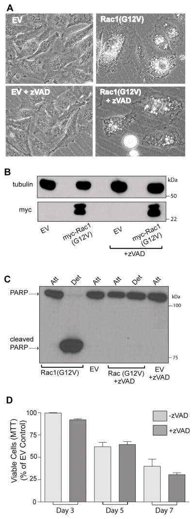
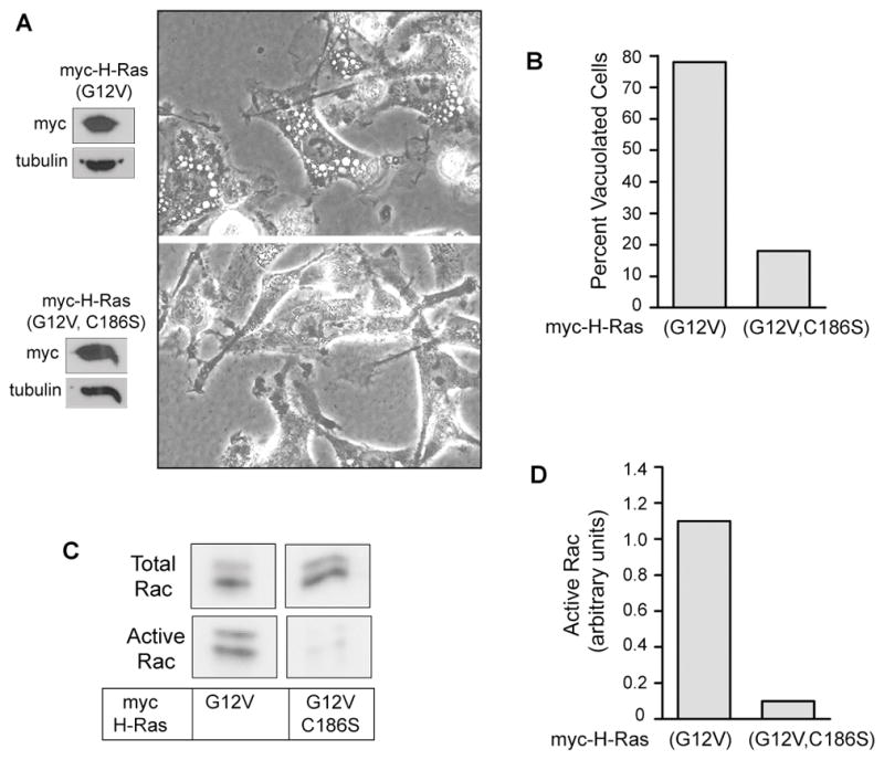
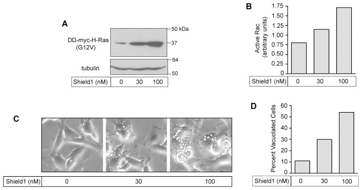
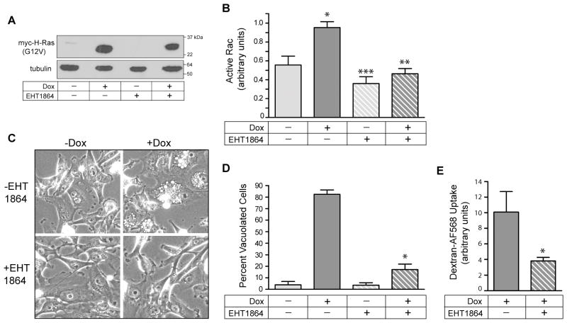
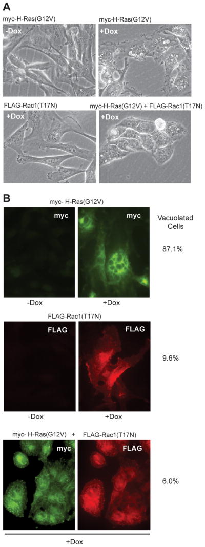
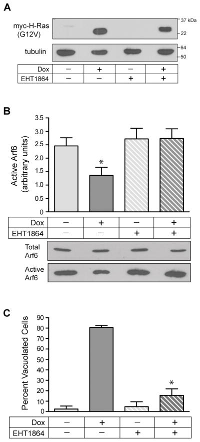
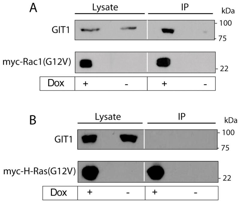
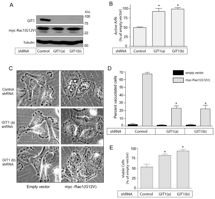
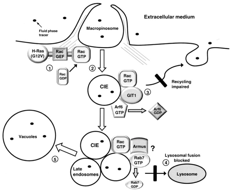
Methuosis is a unique form of nonapoptotic cell death triggered by alterations in the trafficking of clathrin-independent endosomes, ultimately leading to extreme vacuolization and rupture of the cell. Methuosis can be induced in glioblastoma cells by expression of constitutively active Ras. This study identifies the small GTPases, Rac1 and Arf6, and the Arf6 GTPase-activating protein, GIT1, as key downstream components of the signaling pathway underlying Ras-induced methuosis. The extent to which graded expression of active H-Ras(G12V) triggers cytoplasmic vacuolization correlates with the amount of endogenous Rac1 in the active GTP state. Blocking Rac1 activation with the specific Rac inhibitor, EHT 1864, or coexpression of dominant-negative Rac1(T17N), prevents the accumulation of vacuoles induced by H-Ras(G12V). Coincident with Rac1 activation, H-Ras(G12V) causes a decrease in the amount of active Arf6, a GTPase that functions in the recycling of clathrin-independent endosomes. The effect of H-Ras(G12V) on Arf6 is blocked by EHT 1864, indicating that the decrease in Arf6-GTP is directly linked to the activation of Rac1. Constitutively active Rac1(G12V) interacts with GIT1 in immunoprecipitation assays. Ablation of GIT1 by short hairpin RNA prevents the decrease in active Arf6, inhibits vacuolization, and prevents loss of cell viability in cells expressing Rac1(G12V). Together, the results suggest that perturbations of endosome morphology associated with Ras-induced methuosis are due to downstream activation of Rac1 combined with reciprocal inactivation of Arf6. The latter seems to be mediated through Rac1 stimulation of GIT1. Further insights into this pathway could suggest opportunities for the induction of methuosis in cancers that are resistant to apoptotic cell death.


Figures

Figure 1
Figure 1
Expression of activated Rac1 induces caspase-independent death in U251 glioblastoma cells. A) Cells were infected with retrovirus encoding myc-Rac1(G12V) or virus without an cDNA insert (empty vector, EV). On the day after infection, 50 μM z-VAD-fmk was added to half of the cultures, and the others were maintained without the caspase inhibitor. On day-6 after infection A) live cells were examined by phase contrast microscopy, B) western blot analysis was performed to verify expression of myc-Rac1(G12V) and C) attached and detached cells were harvested for analysis of PARP cleavage. There were no detached cells in the EV controls. D) Cells expressing Rac1(G12V) were subjected to MTT assays over a period of 7 days to compare their viability with and without zVAD (the inhibitor was replenished every day). The results shown in the bar graph are from three independent experiments (mean ± SD). The decreases in viable cells at day 3 and day 7 (+/− zVAD) were significant at p 0.04 .
Figure 2
Figure 2
Expression of activated H-Ras causes an increase in the amount of active Rac1 in U251 glioblastoma cells. A) Cells were nucleofected with expression vectors encoding the indicated Ras constructs. After 24 h live cells were examined by phase contrast microscopy and harvested for western blot analysis to check expression of the myc-tagged Ras proteins. B) Cells were scored for vacuolization as described in Materials and Methods. C) Pull-down assays for activated Rac1 were performed on lysates from two pooled 10 cm dishes of cells. The lower blots show the active endogenous Rac1 that bound to the GST-PAK1, and the upper panels show the total Rac1 in an aliquot of the cell lysate. D) The immunoblot signals for active and total Rac1 were quantified and the units for active Rac1 were normalized to total Rac as described in Materials and Methods.
Figure 3
Figure 3
Rac1 activation and cellular vacuolization correlate with increasing levels of H-Ras(G12V) expression in U251 glioblastoma cells. A) Cells were grown in the presence of increasing concentrations of Shield1 to obtain graded expression levels of DD-myc-H-Ras(G12V), using the ProteoTuner system. The blot shows the expressed Ras protein detected with the myc antibody. In a separate blot (not shown) an antibody against H-Ras was used to determine the ratio of expressed DD-myc-H-Ras(G12V) to endogenous H-Ras. The results were 1.4 with no Shield1, 3.0 with 30 nM Shield1, and 4.5 with 100 nM Shield1. B) One day after addition of Shield1, the amount of active endogenous Rac1, normalized to total Rac1, was determined as described in Materials and Methods. C &D) Cell morphology was assessed by phase contrast microscopy and the percentage of cells that were vacuolated was determined.
Figure 4
Figure 4
Blocking Rac1 activation in glioblastoma cells prevents Ras-induced vacuolization. A) The stable cell line, U251-C18, was incubated for 3d in the presence or absence of Dox (1 μg/ml), with or without the Rac1 inhibitor, EHT 1864 (25 μM). Inducible expression of myc-H-Ras(G12V) was verified by western blot analysis. B) Rac activation assays were performed on cells subjected to each condition. The results are the mean (± SD) of three independent determinations performed on separate cultures. The increase in active Rac1 in the cells expressing H-Ras(G12V) (+Dox alone) (*) was significant at p < 0.001compared with the cells not expressing Ras (−Dox). The decrease in active Rac1 in the cultures expressing H-Ras(G12V) in the presence of EHT 1864 (+Doc, +EHT 1864) (**) was significant at p < 0.001 compared with the corresponding control (+ Dox, −EHT 1864). The decrease in the basal level of active Rac1in the −Dox cultures treated with EHT1864 (***) was significant at p <0.05. C&D) At the same time that cultures were harvested for the Rac activation assays, parallel cultures were examined by phase contrast microscopy to determine the percentage of cells that were vacuolated. The results of three separate experiments (mean ± SD) are shown. The suppression of vacuolization by the Rac inhibitor in the cultures expressing H-Ras(G12V) (+Dox, +EHT 1864) (*) was significant at p < 0.0001 compared to the cells expressing H-Ras(G12V) without the Rac inhibitor (+Dox, −EHT 1864). E) Uptake of dextran-AF568 was measured in the +Dox cells expressing myc-H-Ras(G12V), with or without EHT1864. The results are the mean ± S.D. of separate determinations on three cultures. The decrease in dextran uptake in the cells treated with the Rac inhibitor (*) was significant at p < 0.04.
Figure 5
Figure 5
Expression of dominant-negative Rac1 blocks the induction of vacuolization by active H-Ras. U251 cell lines capable of conditional expression of myc-H-Ras(G12V), FLAG-Rac1(T17N) or both constructs, were generated using the pRetroX-Tight-Pur Tet-On system as described in Materials and Methods. A) Phase contrast microscopy of the cells after 2 days with or without Dox in the medium. B) Immunofluorescence microscopy demonstrates expression of myc-H-Ras(G12V) and/or FLAG-Rac1(T17N) only in the presence of Dox. The percentage of vacuolated cells in each culture was determined by scoring only the cells with visible expression of the myc and/or FLAG constructs. The results are based on counting a minimum of 70 cells.
Figure 6
Figure 6
Suppression of Tiam1 or Eps8 expression does not prevent Ras-induced vacuolization of glioblastoma cells. A) shRNA-mediated knockdown of Tiam1 or Eps8 expression was performed as described in Materials and Methods. Western blots of whole cell lysates show that the specific shRNAs reduced expression of Tiam1 or Eps8 by >90% compared with the respective controls. The loading controls, α-tubulin and LDH, confirm that comparable amounts of cell protein were applied to the lanes. B) When cells were grown in the presence of Dox to induce expression of myc-H-Ras(G12V) (lower blots), there was no decrease in vacuolization of the cells lacking Tiam1 or Eps8, compared with the control. Vacuolization was assessed by phase contrast microscopy and quantified as described in Materials and Methods.
Figure 7
Figure 7
Expression of activated H-Ras causes a Rac1-dependent decrease in the amount of active Arf6, which is essential for vacuolization of glioblastoma cells. A) U251-C18 cells were incubated for 3d in the presence or absence of Dox (1 μg/ml), with or without the Rac1 inhibitor, EHT 1864 (25 μM). Inducible expression of myc-H-Ras(G12V) was verified by western blot analysis. B) Arf6 activation assays were performed on cells subjected to each condition, with active Arf6 normalized to total Arf6 as described in Materials and Methods. The results are the mean (± SD) of three independent determinations performed on separate cultures. The decrease in active Arf6 in the cells expressing H-Ras(G12V) (+Dox alone) (*) was significant at p < 0.01compared with the cells not expressing Ras (−Dox) or the cells incubated with the Rac inhibitor, EHT 1864. Representative immunoblots of the raw Arf-6 GTP pull-downs are shown below the bar graph. C) At the time that cultures were harvested for the Arf6 activation assays, parallel cultures were examined by phase contrast microscopy to determine the percentage of cells that were vacuolated. The results of three separate experiments (mean ± SD) are shown. The suppression of vacuolization caused by addition of the Rac inhibitor to cultures expressing H-Ras(G12V) (+Dox, +EHT 1864) (*) was significant at P < 0.0001 compared to the cells expressing H-Ras(G12V) without the Rac inhibitor (+Dox, −EHT 1864).
Figure 8
Figure 8
Active Rac1 interacts with the Arf6 GAP, GIT1. A) Three days after adding Dox to induce expression of myc-Rac1(G12V) in a stable U251 cell line, cells were lysed and the myc-tagged protein was collected on agarose beads conjugated with anti myc antibody. Samples of whole-cell lysate and proteins eluted from the myc-agarose beads were subjected to western blot analysis with monoclonal antibodies against GIT1 or myc. Immunoprecipitation of lysate from cells that did not receive Dox was performed to control for non-specific precipitation of GIT1. B) The same experiment was performed with stable U251 cells induced to express myc-H-Ras(G12V) to establish that GIT1 does not associate with activated H-Ras.
Figure 9
Figure 9
Suppression of GIT1 expression prevents the Rac1(G12V)-mediated decrease in active Arf6 and protects glioblastoma cells from methuosis. Stable cell lines were generated from U251 cells infected with lentivirus encoding shRNA against GFP (control) and two different shRNAs against GIT1 (GIT1 a & b). A) The control and GIT1 knockdown cell lines were infected with retrovirus encoding myc-Rac1(G12V), and three days after infection the cells were subjected to western blot analysis to check expression of myc-Rac1(G12V) and GIT1. B) On the fourth day after infection the control and GIT1 knockdown cell lines were assayed for the amount of active Arf6 as described in Materials and Methods. The results show the amount of active Arf6 in the cells expressing myc-Rac1(G12V), expressed as percent of the value obtained for the same cell line infected with empty retroviral vector (mean + SD for three experiments). The differences between each of the GIT1 knockdown cell lines and the control cell line were significant at p<0.004. C & D) On the fourth day after infection with empty or myc-Rac1(G12V) retroviral vectors, the control and GIT1 knockdown cell lines were examined by phase contrast microscopy to determine the percentage of cells that were vacuolated. The results of three separate experiments (mean ± SD) are shown in the bar graph. The suppression of Rac1-induced vacuolization in the GIT1 knockdown cell lines was significant at p < 0.0001 compared to the control cell line. E) On the fifth day after infection with empty or myc-Rac1(G12V) lentiviral vectors, the control and GIT1 knockdown cells were assayed for cell viability using MTT. Assays were performed on quadruplicate cultures in a 96-well plate, and the results for the cells infected with myc-Rac1(G12V) retro virus were expressed as percent of the same cells infected with empty vector. The increased viability observed in each of the GIT1 knockdown cell lines was significant at p<0.007 relative to the shRNA control cell line.
Figure 10
Figure 10
A hypothetical model for Ras-induced methuosis suggested by the present findings: 1. Expression of constitutively activated H-Ras reaches a threshold sufficient to stimulate the activity of an unidentified Rac1 guanine nucleotide exchange factor (GEF). 2. The resulting increase in the pool of active Rac1 enhances macropinocytosis. 3. As the pool of active Rac1 increases beyond normal physiological levels, Rac1-GTP associates with GIT1 and stimulates Arf6 GAP activity. The consequent decrease in the pool of active Arf6 impairs recycling of clathrin-independent endosomes (CIE). 4. CIEs derived from macropinosomes acquire some characteristics of late endosomes (Rab7, LAMP1), but fail to merge with lysosomes, due to trafficking defects at the late endosome/lysosome boundary. One possible mechanism for a block in lysosomal fusion could be Rac1 stimulation of the Rab7 GAP, Armus, resulting in inactivation of Rab7, as recently described by Frasa et al. (66). 5. The accumulated CIE and late endosomal vesicles coalesce to form progressively larger vacuolar structures, which ultimately displace most of the cytoplasm and lead to cell detachment from the substratum and disruption of cell membrane integrity.

Similar articles

Cited by

References

    1. Bursch W, Ellinger A, Gerner C, Frohwein U, Schulte-Hermann R. Programmed cell death (PCD). Apoptosis, autophagic PCD, or others? Ann N Y Acad Sci. 2000;926:1–12. - PubMed
    1. Lockshin RA, Zakeri Z. Apoptosis, autophagy, and more. Int J Biochem Cell Biol. 2004;36:2405–2419. - PubMed
    1. Gozuacik D, Kimchi A. Autophagy as a cell death and tumor suppressor mechanism. Oncogene. 2004;23:2891–2906. - PubMed
    1. Sperandio S, de Belle I, Bredesen DE. An alternative, nonapoptotic form of programmed cell death. Proc Natl Acad Sci U S A. 2000;97:14376–14381. - PMC - PubMed
    1. Wang Y, Li X, Wang L, Ding P, Zhang Y, Han W, Ma D. An alternative form of paraptosis-like cell death, triggered by TAJ/TROY and enhanced by PDCD5 overexpression. J Cell Sci. 2004;117:1525–1532. - PubMed

Publication types

MeSH terms