. 2011 Mar;21(3):636-46.
doi: 10.1093/cercor/bhq137. Epub 2010 Aug 16.

Puberty influences medial temporal lobe and cortical gray matter maturation differently in boys than girls matched for sexual maturity

Affiliations

Puberty influences medial temporal lobe and cortical gray matter maturation differently in boys than girls matched for sexual maturity

Jennifer E Bramen et al. Cereb Cortex. 2011 Mar.

Abstract

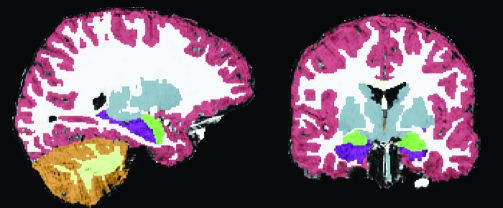
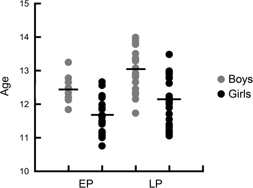
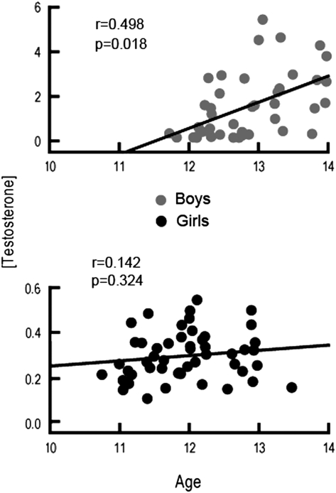
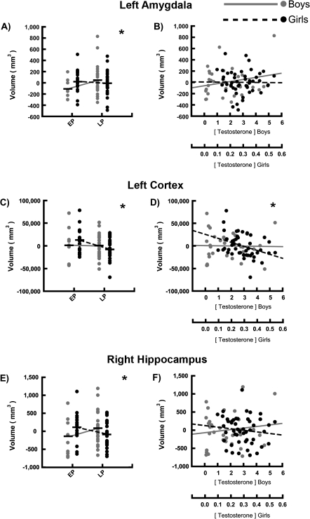
Sex differences in age- and puberty-related maturation of human brain structure have been observed in typically developing age-matched boys and girls. Because girls mature 1-2 years earlier than boys, the present study aimed at assessing sex differences in brain structure by studying 80 adolescent boys and girls matched on sexual maturity, rather than age. We evaluated pubertal influences on medial temporal lobe (MTL), thalamic, caudate, and cortical gray matter volumes utilizing structural magnetic resonance imaging and 2 measures of pubertal status: physical sexual maturity and circulating testosterone. As predicted, significant interactions between sex and the effect of puberty were observed in regions with high sex steroid hormone receptor densities; sex differences in the right hippocampus, bilateral amygdala, and cortical gray matter were greater in more sexually mature adolescents. Within sex, we found larger volumes in MTL structures in more sexually mature boys, whereas smaller volumes were observed in more sexually mature girls. Our results demonstrate puberty-related maturation of the hippocampus, amygdala, and cortical gray matter that is not confounded by age, and is different for girls and boys, which may contribute to differences in social and cognitive development during adolescence, and lasting sexual dimorphisms in the adult brain.


Figures

Figure 1.
Figure 1.
Example segmented brain. On the left is a sagittal view and on the right is a coronal view demonstrating tissue segmentation of a single participant’s brain. Segmentation includes cortical gray (red) and white matter (white) segmentation, and subcortical structures, the majority of which are colored in gray. Subcortical regions of interest are highlighted in bright colors (the hippocampus colored in purple and the amygdala in green).
Figure 2.
Figure 2.
Physical sexual maturity as a function of age in boys and girls. Scatter plot showing the relationship betweens between sexual maturity (x-axis) and age (y-axis) in boys and girls. While the y-axis is in years, age is actually plotted in months, to best demonstrate the age of individual adolescents as well as the narrowness of the age range studied. Sexual maturity was assessed using TS based on physical exam. Pre/early puberty (EP) represented TS 1 or 2, while mid/late puberty (LP) represented TS 3–5. The mean for each sex/sexual maturity group is represented using a horizontal bar through the corresponding scatter plot. While there was a correlation between sexual maturity and age for both boys and girls, there was no significant interaction between sex, age, and sexual maturity. Therefore, age was not a confounding factor when testing sex by sexual maturity interactions on brain volumes in this sample.
Figure 3.
Figure 3.
Circulating TES levels as a function of age. Scatter plots showing the relationships between circulating TES (y-axis) and age (x-axis) in boys and girls. As with Figure 2, age is plotted in months. Circulating TES level was an order of magnitude greater in boys than in girls (reflected in the range on the y-axis). The correlation coefficient (r) is also displayed. TES was significantly correlated with age in boys, but not in girls.
Figure 4.
Figure 4.
Interactions between sex and the effect of pubertal status on development of select regions of interest. Sex differences in the effects of puberty on regional volume were assessed and scatter plots demonstrate sex by pubertal status (x axis) on gray matter volume (y axis) interactions in the left amygdala, left cortex, and right hippocampus. Two measures of pubertal status are plotted—(left) sexual maturity, measured with TS using TS (Tanner breast/genital exam) and converted to a 2-level variable: pre/early puberty (TS = 1–2) (EP) and mid/late puberty (TS = 3–5) (LP); and (right) circulating TES, with respect to regional volumes. The mean is represented using a horizontal black bar through the corresponding scatter plot for binarized measures of TS for each group (A, C, and E). A line connecting mean regional volumes of adolescents in EP and LP is drawn separately in boys and girls to demonstrate the interactions found. Circulating TES was plotted against regional volume in boys and girls (B, D, and F). Significant interactions between sex and the effect of sexual maturity on regional volume were found (A, C, and E). Significant interactions between sex and the effect of circulating TES on regional volume were found (D). No significant interactions between sex and the effect of sexual maturity on regional volume were found in B or F. *Significance threshold was P < 0.05 for sex by pubertal status effect on volume interaction.

Similar articles

Cited by

References

    1. Ahmed EI, Zehr JL, Schulz KM, Lorenz BH, DonCarlos LL, Sisk CL. Pubertal hormones modulate the addition of new cells to sexually dimorphic brain regions. Nat Neurosci. 2008;11:995–997. - PMC - PubMed
    1. Aleman A, Swart M. Sex differences in neural activation to facial expressions denoting contempt and disgust. Plos ONE. 2008;3:e3622. - PMC - PubMed
    1. Angold A, Costello EJ, Erkanli A, Worthman CM. Pubertal changes in hormone levels and depression in girls. Psychol Med. 1999;29:1043–1053. - PubMed
    1. Archer J. Rodent sex differences in emotional and related behavior. Behav Biol. 1975;14:451–479. - PubMed
    1. Arnold AP. Sex chromosomes and brain gender. Nat Rev Neurosci. 2004;5:701–708. - PubMed

Publication types