Review
. 2010 Aug 15;24(16):1680-94.
doi: 10.1101/gad.1955310.

Expanded roles of the Fanconi anemia pathway in preserving genomic stability

Affiliations
Review

Expanded roles of the Fanconi anemia pathway in preserving genomic stability

Younghoon Kee et al. Genes Dev. .

Abstract

Studying rare human genetic diseases often leads to a better understanding of normal cellular functions. Fanconi anemia (FA), for example, has elucidated a novel DNA repair mechanism required for maintaining genomic stability and preventing cancer. The FA pathway, an essential tumor-suppressive pathway, is required for protecting the human genome from a specific type of DNA damage; namely, DNA interstrand cross-links (ICLs). In this review, we discuss the recent progress in the study of the FA pathway, such as the identification of new FANCM-binding partners and the identification of RAD51C and FAN1 (Fanconi-associated nuclease 1) as new FA pathway-related proteins. We also focus on the role of the FA pathway as a potential regulator of DNA repair choices in response to double-strand breaks, and its novel functions during the mitotic phase of the cell cycle.


Figures
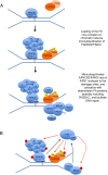
Figure 1.
Figure 1.
A schematic model for the FA pathway. (A) Activation of the FA pathway. DNA ICL is directly recognized by FANCM–FAAP24–MHF protein complex. This complex recruits the FA core complex by direct interaction between FANCM and FANCF. The recruited FA core complex, containing a PHD E3 ubiquitin ligase domain in the FANCL subunit, subsequently monoubiquitinates its two substrates, FANCD2 and FANCI, on chromatin. The monoubiquitinated FANCD2–FANCI becomes an active form, recruits newly identified nuclease FAN1 or interacts with a series of DNA repair proteins (including BRCA1, PALB2 [FANCN], BRCA2, and FANCJ [BACH1/BRIP1]) at the damaged sites, and facilitates downstream repair pathways. RAD51C, a newly identified FA-like protein, may have a functional interaction with FANCD2 at this step. FANCD2–FANCI probably also recruits other nucleases and TLS polymerases to process the ICL (not shown). The new players in the FA pathway described in the text are highlighted in dashed lines. (B) The FA pathway cross-talks with ATR–CHK1 checkpoint proteins. ATR and its effector kinase, CHK1, are required for damage-inducible activation of the FA pathway. ATR–CHK1 phosphorylates (red arrows) multiple FA and FA-associated proteins, including FANCA, FANCE, FANCD2, FANCI, and BRCA1. FANCM is also phosphorylated upon DNA damage by an unknown kinase. In turn, the stability and activity of ATR–CHK1 are promoted (black arrows) by the FANCM–FAAP24 heterodimer and FANCJ by independent mechanisms. FANCM also mediates the formation of the BRAFT complex, which contains the FA core complex members and the BLM complex containing Topo IIIα and RMI1/2 (not shown).
Figure 2.
Figure 2.
A schematic for DSB repair by HR and NHEJ pathways. DSBs can be repaired by either HR or NHEJ. New players (RAD51C and POLN/HEL308) in the HR pathway are displayed in the left side of the figure, and are described in detail below. For initiation of HR, DSB ends must be resected to expose 3′ overhangs of ssDNA by the exonuclease activity of CtIP. The exposed ssDNA is rapidly coated with RPA. RPA is then replaced by RAD51, the step facilitated by BRCA1, PALB2, and BRCA1. A mediator protein, RAD52, also helps RAD51 loading (not shown). The resulting ssDNA–RAD51 presynaptic filaments are capable of invading the homologous region in the nearby duplex DNA, forming a triplex DNA called a D-loop. DNA polymerases further extend DNA synthesis (possibly by combined or redundant activities of POLη, POLδ, and POLN), and the recombination intermediates are finally resolved to complete the repair (not shown). RAD51C, one of the five RAD51 paralogs found in human cells, appears to promote loading of RAD51 (required for RAD51 foci formation) at an early step of HR. RAD51C—by forming a complex with another paralog, XRCC3—may also act to resolve Holliday junctions at the later step of HR (Liu et al. 2007). NHEJ directly seals two DSB ends and does not generally require DSB end resection. Binding of Ku70–80 heterodimer (the regulatory subunits of DNA-PK) at DSB ends recruits DNA-PKcs. The activated DNA-PKcs recruits DNA ligase IV (LIG4), which subsequently joins two broken DNA ends. NHEJ can occur without homology, such as ligation between two blunt ends or ends with overhangs that can be processed by resection or fill-in. Recent studies suggested that the MRE11 nuclease may function in end processing (Zha et al. 2009) A minor form of NHEJ, microhomology-directed NHEJ, is not described here to keep simplicity. In addition to these “core” NHEJ proteins, other factors, such as MRE11 of the MRN complex, regulate certain types of NHEJ (Deng et al. 2009; Zha et al. 2009).
Figure 3.
Figure 3.
A simplified scheme for ICL repair. Progression of replication forks is blocked by ICL. The stalled replication forks can trigger multiple surveillance mechanisms, one of them being monobiquitination of the FANCD2/I heterodimer. The initial event is thought to be the incising of ICLs by serial or combined activities of XPF–ERCC1 and MUS81–EME1. Potentially, the newly identified FAN1 might act on this step. These nucleases cut one side of the damaged DNA, unhooking the ICL and leaving a gap. The gap is subsequently bypassed by TLS polymerases, probably REV1, followed by removal of the monoadducts and repairing the gap. DSBs, a byproduct of the ICL repair process, are subsequently repaired by HR (see Fig. 2). Activated FANCD2/FANCI (brown circle) were shown to be required at multiple steps, including the nucleolytic incision and the TLS-mediated bypass (Knipscheer et al. 2009). Whether FANCD2/FANCI also functions directly in the HR process is unknown.
Figure 4.
Figure 4.
A hypothetical model for NHEJ-suppressive mechanisms during DSB repair. For repairing “replication-associated” DSBs, multiple mechanisms can be employed to promote HR. (Left panel) BRCA1 may have a function to displace 53BP1 from DSB ends, allowing ATM-CtIP-dependent DNA end resection and initiation of HR. In BRCA1-deficient cells, sustained activity of 53BP1 at DSB ends may inhibit the end resection, while it facilitates recruitment of NHEJ factors. (Right panel) FA proteins (FANCD2, FANCI, BRCA2, PALB2, or BRCA1) may have roles in precluding NHEJ factors (e.g., Ku70/80 and DNA-PKcs) from binding to DSB ends by either direct inhibition or facilitating HR initiating events, such as DNA end resection. Deletion or pharmacological inhibition of the NHEJ pathway rescues FA-deficient phenotypes.

Similar articles

Cited by

References

    1. Adamo A, Collis SJ, Adelman CA, Silva N, Horejsi Z, Ward JD, Martinez-Perez E, Boulton SJ, La Volpe A 2010. Preventing nonhomologous end joining suppresses DNA repair defects of Fanconi Anemia. Mol Cell. 39: 25–35 - PubMed
    1. Ali AM, Singh TR, Meetei AR 2009. FANCM–FAAP24 and FANCJ: FA proteins that metabolize DNA. Mutat Res 668: 20–26 - PMC - PubMed
    1. Andreassen PR, D'Andrea AD, Taniguchi T 2004. ATR couples FANCD2 monoubiquitination to the DNA-damage response. Genes Dev 18: 1958–1963 - PMC - PubMed
    1. Arlt MF, Xu B, Durkin SG, Casper AM, Kastan MB, Glover TW 2004. BRCA1 is required for common-fragile-site stability via its G2/M checkpoint function. Mol Cell Biol 24: 6701–6709 - PMC - PubMed
    1. Bakker ST, van de Vrugt HJ, Rooimans MA, Oostra AB, Steltenpool J, Delzenne-Goette E, van der Wal A, van der Valk M, Joenje H, te Riele H, et al. 2009. Fancm-deficient mice reveal unique features of Fanconi anemia complementation group M. Hum Mol Genet 18: 3484–3495 - PubMed

Publication types