. 2010 Aug 15;24(16):1709-17.
doi: 10.1101/gad.1958410.

Constitutively active NF-kappaB triggers systemic TNFalpha-dependent inflammation and localized TNFalpha-independent inflammatory disease

Affiliations

Constitutively active NF-kappaB triggers systemic TNFalpha-dependent inflammation and localized TNFalpha-independent inflammatory disease

Jie Dong et al. Genes Dev. .

Abstract

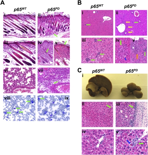
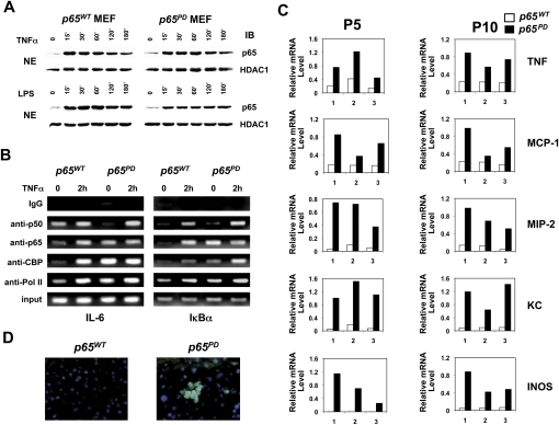
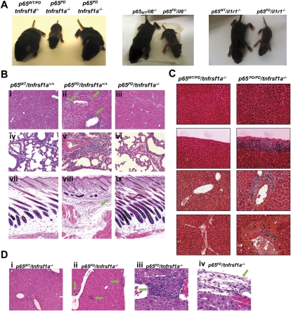
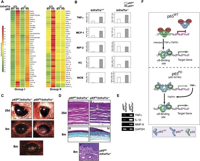
NF-kappaB is well established as a key component of the inflammatory response. However, the precise mechanisms through which NF-kappaB activation contributes to inflammatory disease states remain poorly defined. To test the role of NF-kappaB in inflammation, we created a knock-in mouse that expresses a constitutively active form of NF-kappaB p65 dimers. These mice are born at normal Mendelian ratios, but display a progressive, systemic hyperinflammatory condition that results in severe runting and, typically, death 8-20 d after birth. Examination of homozygous knock-in mice demonstrates significant increases in proinflammatory cytokines and chemokines. Remarkably, crossing this strain with mice lacking TNF receptor 1 (TNFR1) leads to a complete rescue of the hyperinflammatory phenotype. However, upon aging, these rescued mice begin to display chronic keratitis accompanied by increased corneal expression of TNFalpha, IL-1beta, and MMP-9, similar to that seen in human keratoconjunctivitis sicca (KCS) or "dry eyes." Therefore, our results show that, while constitutively active NF-kappaB can trigger systemic inflammation, it does so indirectly, through increased TNF production. However, certain inflammatory disease states, such as keratitis or KCS, a condition that is seen in Sjogren's syndrome, are dependent on NF-kappaB, but are independent of TNFR1 signaling.


Figures

Figure 1.
Figure 1.
Generation and phenotypic characterization of p65 S276D (PD) knock-in mice. (A) PCR analysis of mouse tail DNA. Restriction enzyme digestion with EagI was performed to detect the PD mutation on p65. (B) Sequencing of p65 cDNA obtained by RT–PCR from wild-type and PD/PD homozygous mice. (C) Immunoblot analysis of p65 expression levels in wild-type and PD homozygous MEFs generated from embryos at 12.5 dpc. (D) Interaction between p65 and IκBα or IκBβ detected by immunoprecipitation in MEFs. (E) Gross appearance of wild-type and PD/PD homozygous knock-in P10 mice.
Figure 2.
Figure 2.
Characterization of hyperinflammatory phenotype in PD mice. (A) Hematoxylin and eosin (H&E) staining of skin (panels i–v) and lung (panels vi,vii), and peripheral blood smear analysis (panels viii,ix) of wild-type and PD/PD littermates at P10. Bars: panels i,ii, 50 μm; panels iiivii, 20 μm; panels viii,ix, 10 μm. (B) H&E staining of wild-type and PD/PD livers at P10. Bars: panels i,ii, 50 μm; panels iiiv, 20 μm. (C) Gross appearance and H&E staining of wild-type and PD/PD livers at 3.5 mo. Bars: panels ii,iii, 50 μm; panlesl iv,v, 20 μm.
Figure 3.
Figure 3.
Hyperactivation of NF-κB induced by PD mutation. (A) Translocation of p65 following TNFα (10 ng/mL) or LPS (10 μg/mL) stimulation in MEFs. (B) ChIP assay was performed with untreated or TNFα-treated (2 h) MEFs using the indicated antibodies. Precipitated IL-6 κB site DNA and IκBα κB site DNA were assayed by semiquantitative PCR. (C) Differential expression of NF-κB-regulated genes in wild-type and PD/PD livers at P5 and P10. Total RNA isolated from three pairs of wild-type and PD/PD littermates was quantified by real-time RT–PCR, and was normalized to the level of GAPDH. (D) Immunostaining of TNF in wild-type and PD/PD livers at P10. Liver cross-sections were immunostained with FITC-conjugated antibody recognizing TNF. Nuclei were stained by DAPI.
Figure 4.
Figure 4.
Phenotype of PD homozygous knock-in mice rescued by knocking out TNFR1. (A) Phenotype of PD/PD mice can be rescued by crossing with TNFR1 knockouts, but not IL6 or IL1R knockouts. Gross appearance of P10 littermates is presented. (B) H&E staining of major organs to confirm the rescue phenomenon resulted from knocking out TNFR1. Organs from P10 littermates were analyzed. Bars: panels iiii,viiix, 50 μm; panels ivvi, 20 μm. (C) H&E staining of liver in aged wild-type and PD/PD mice that lack TNFR1. Bars: panels iiii, 50 μm; panels ivvi, 20 μm. (D) H&E staining of the liver in one age-matched wild-type and three PD/PD TNFR1−/− mice that died at 6 mo of age.
Figure 5.
Figure 5.
Regulation of gene expression and KC by p65 PD. (A) Comparison of gene expression in wild-type versus PD/PD livers obtained from P5 mice, either with or without TNFR1, using Affymetrix microarrays. Representative genes in two groups of genes were hierarchically clustered and displayed. Each row represents a single gene, and each column represents an experimental sample. The ratio of the abundance of transcripts of each gene to the median abundance of the gene's transcript among all the samples is represented by the color in the matrix, as indicated. Relative expression levels are shown at the bottom. (B) Total RNA isolated from the livers of three pairs of wild-type p65 and PD/PD littermates, either with or without TNFR1, at P5 was quantified by real-time RT–PCR, and was normalized to the level of GAPDH. The average mRNA level of three mice in each group is presented. Ocular lesions accompanying inflammation developed in the eye of the PD homozygous knock-in mice rescued by knocking out TNFR1. (C) Gross appearance of cornea in weanling (25d) and adult (8m) mice. Bar: panels i–v, 1 mm. (D) Corneal histology in weanling (25d; H&E staining in panels i,ii) and adult (8m; masson's trichrome staining in panels iii,iv; H&E staining in panel v) mice. Bars: panels i,ii, 20 μm; panels iii,iv, 100 μm; panel v, 50 μm. (E) Corneal expression of TNFα, IL-1β, and MMP-9 at 25 d, determined by semiquantitative RT–PCR. (F) A model of the mechanism through which p65 S276D mutant protein hyperactivates gene expression. Expression of the PD mutant protein creates a constitutively active form of NF-κB (consisting of p65 homodimers) that up-regulates TNFα, which then acts in a paracrine manner through TNFR1 to trigger systemic hyperinflammation.

References

    1. Adler AS, Sinha S, Kawahara TL, Zhang JY, Segal E, Chang HY 2007. Motif module map reveals enforcement of aging by continual NF-κB activity. Genes Dev 21: 3244–3257 - PMC - PubMed
    1. Anest V, Cogswell PC, Baldwin AS Jr 2004. IκB kinase α and p65/RelA contribute to optimal epidermal growth factor-induced c-fos gene expression independent of IκBα degradation. J Biol Chem 279: 31183–31189 - PubMed
    1. Chen LF, Greene WC 2004. Shaping the nuclear action of NF-κB. Nat Rev Mol Cell Biol 5: 392–401 - PubMed
    1. Chen LF, Williams SA, Mu Y, Nakano H, Duerr JM, Buckbinder L, Greene WC 2005. NF-κB RelA phosphorylation regulates RelA acetylation. Mol Cell Biol 25: 7966–7975 - PMC - PubMed
    1. Deak M, Clifton AD, Lucocq LM, Alessi DR 1998. Mitogen- and stress-activated protein kinase-1 (MSK1) is directly activated by MAPK and SAPK2/p38, and may mediate activation of CREB. EMBO J 17: 4426–4441 - PMC - PubMed

Publication types

LinkOut - more resources