. 2010 Aug 15;24(16):1772-86.
doi: 10.1101/gad.1954910.

Systematic screen reveals new functional dynamics of histones H3 and H4 during gametogenesis

Affiliations

Systematic screen reveals new functional dynamics of histones H3 and H4 during gametogenesis

Jérôme Govin et al. Genes Dev. .

Abstract

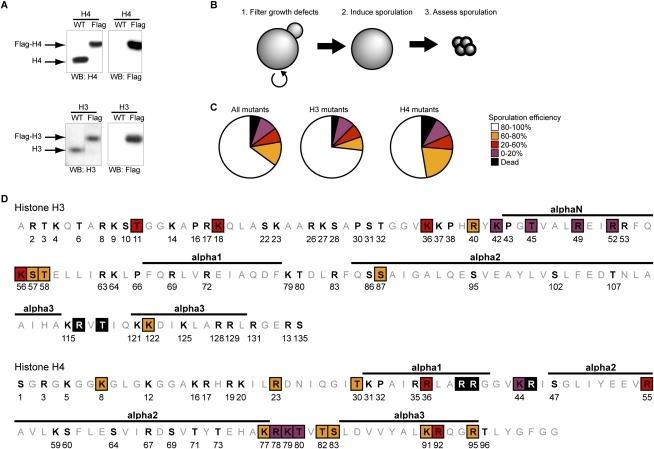
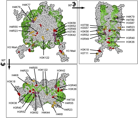
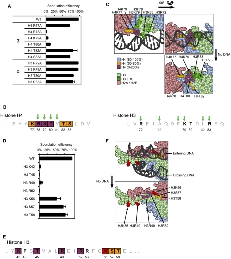
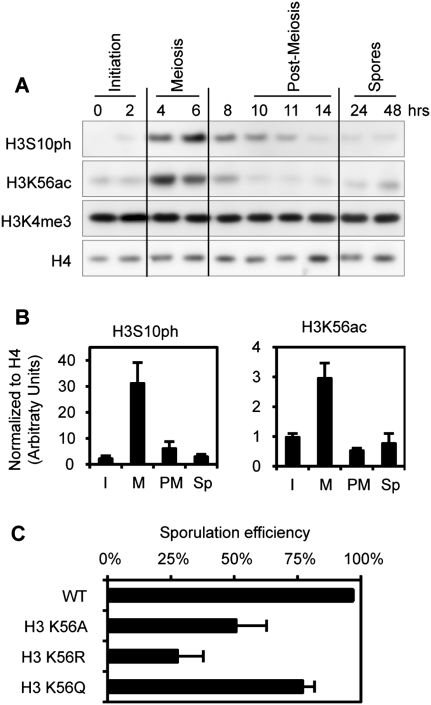
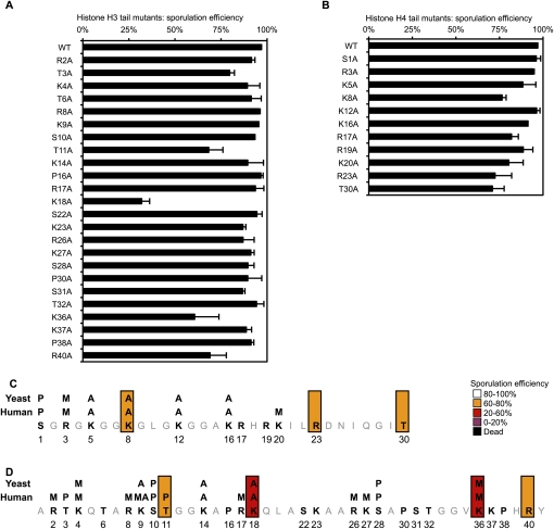
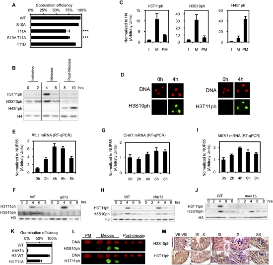
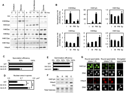
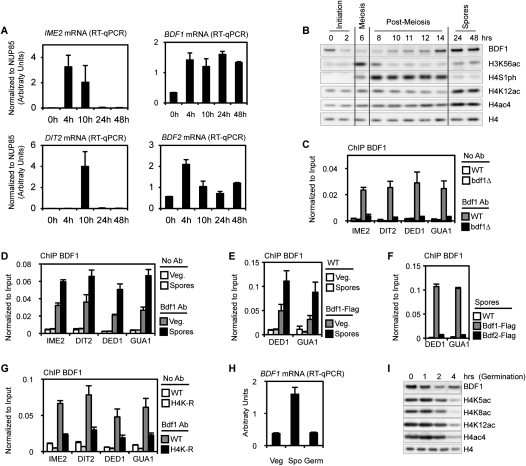
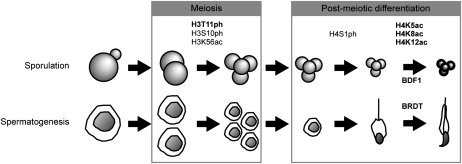
Profound epigenetic differences exist between genomes derived from male and female gametes; however, the nature of these changes remains largely unknown. We undertook a systematic investigation of chromatin reorganization during gametogenesis, using the model eukaryote Saccharomyces cerevisiae to examine sporulation, which has strong similarities with higher eukaryotic spermatogenesis. We established a mutational screen of histones H3 and H4 to uncover substitutions that reduce sporulation efficiency. We discovered two patches of residues-one on H3 and a second on H4-that are crucial for sporulation but not critical for mitotic growth, and likely comprise interactive nucleosomal surfaces. Furthermore, we identified novel histone post-translational modifications that mark the chromatin reorganization process during sporulation. First, phosphorylation of H3T11 appears to be a key modification during meiosis, and requires the meiotic-specific kinase Mek1. Second, H4 undergoes amino tail acetylation at Lys 5, Lys 8, and Lys 12, and these are synergistically important for post-meiotic chromatin compaction, occurring subsequent to the post-meiotic transient peak in phosphorylation at H4S1, and crucial for recruitment of Bdf1, a bromodomain protein, to chromatin in mature spores. Strikingly, the presence and temporal succession of the new H3 and H4 modifications are detected during mouse spermatogenesis, indicating that they are conserved through evolution. Thus, our results show that investigation of gametogenesis in yeast provides novel insights into chromatin dynamics, which are potentially relevant to epigenetic modulation of the mammalian process.


Figures

Figure 1.
Figure 1.
A systematic screen identifies H3 and H4 residues essential for sporulation completion. (A) Creation and validation of a histone shuffle strain in the SK1 background. The strain was validated by replacing a wild-type (WT) plasmid with a plasmid expressing Flag-tagged H3 or H4. Detection of H3 and H4 by Western blot confirms that neither genomic copy of the H3 and H4 genes remains, and that the entire population of H3 and H4 is expressed from the plasmid. (B) Schematic of the rationale used for the screen. More than 100 substitution mutants have been created and validated by sequencing. Several substitution mutations were lethal, and were not analyzed further. Sporulation was induced and spore formation was quantified in all other mutants. (C) Statistical analysis of the screen results. The severity of the sporulation defects is described using the following color code: a sporulation efficiency included within 0%–20%, 20%–60%, 60%–80%, and 80%–100% is represented in purple, red, orange, and white, respectively. Lethal mutations are represented in black. This same color-coding is used throughout the figures. (D) Graphic representation of the substitution mutants affected in sporulation. The histone fold is represented at the top of each histone, and is based on data from White et al. (2001).
Figure 2.
Figure 2.
Representation of the nucleosome, highlighting the residues essential for completion of sporulation. The same color-coding is used as in Figure 1. Front and two different lateral views are presented. PDB file 1KX5 was used (Davey et al. 2002), and was color-coded using Accelrys Discovery Studio Visualizer 2.5.
Figure 3.
Figure 3.
Detailed analysis of the LOS and DEE patches. (A) Sporulation efficiency data for the LOS substitution mutants. (B) Graphical representation of the LOS (H4) and the H3 LRS regions. Green arrows represent identified LRS mutations (Park et al. 2002). (C) Three-dimensional representation of the LOS patch. The severity of the sporulation phenotype for each substitution is represented following the same color-coding as in Figure 1C. The H3 LRS mutants are represented in dark green. H2A/H2B, H3, and H4 are represented in light pink, light green, and light blue, respectively. (D) Sporulation efficiency data for DEE mutants. (E) Graphical representation of the DEE patch. (C) Three-dimensional representation of the DEE patch. Refer to C and Figure 1C for details on the color-coding.
Figure 4.
Figure 4.
Dynamics of meiosis-specific histone modifications during sporulation. (A) H3S10ph and H3K56ac were detected by Western blot during sporulation. (B) Quantification of the Western blots, including images presented in A. The time course has been broken into stages, as indicated in A. (I) Initiation; (M) meiosis; (PM) post-meiosis; (Sp) spores. Quantification was performed using two to three biological independent replicates (depending on the time points). (C) Sporulation efficiency data for different H3K56 mutants. H3K56R mimics the unmodified form of H3K56, while H3K56Q mimics the acetylated form of H3K56.
Figure 5.
Figure 5.
Analysis of mutants located in the N-terminal tails of H3 and H4. (A,B) Detailed sporulation efficiency data for histones H3 and H4. (C,D) Graphical representation of the residues essential for sporulation completion (same color-coding as in Fig. 1).
Figure 6.
Figure 6.
H3T11ph is a new meiotic mark in yeast and mammals. (A) Sporulation efficiency data for selected mutants. P-values are 10−7 and 10−3 for H3T11A and H3S10AT11A mutants, respectively (asterisks). (B) Detection of H3T11ph during sporulation by Western blot. (C) Quantification of the Western blots, including images presented in B. The time course has been broken into stages, as indicated in B. (I) Initiation; (M) meiosis; (PM) post-meiosis. Quantification was performed using two to three biological independent replicates (depending on the time points). (D) In situ detection of H3S10ph and H3T11ph by immunofluorescence during sporulation. (E,G,I) Quantifications of mRNA abundance of IPL1, CHK1, and MEK1, respectively. Data were obtained from four independent biological replicates, and normalized to NUP85 (see the Materials and Methods). (F) Ipl1 is essential for H3S10ph deposition. Depletion of Ipl1 during meiosis (using a conditional strain; see the text for details) leads to a loss of H3S10ph, but does not affect H3T11ph. Western blot analysis using indicated antibodies. (H) Chk1 is not required for H3S10ph or H3T11ph deposition. Western analysis of the histone modifications in a chk1Δ strain compared with the wild-type strain. (J) Mek1 is essential for H3T11ph deposition. Western analysis of the histone modifications in a mek1Δ strain compared with the wild-type strain. (K) Germination efficiency. Spores were dissected and then germinated on YPD plates. More than 50 spores were analyzed using two independent isolates of each strain. (L) Analyses of H3S10ph and H3T11ph during mouse spermatogenesis using immunofluorescence. (M) Immunohistochemistry using H3S10ph and H3T11ph antibodies during mouse spermatogenesis.
Figure 7.
Figure 7.
Dynamics of H4 modifications during post-meiotic maturation of the gamete. (A) The phosphorylation and acetylation status of H4 during sporulation analyzed by Western blot. The H3K56ac and H4S1ph pattern confirms that the cells are sporulating synchronously. (B) Quantification of the Western blots, including images presented in A. Quantification was performed using two to three biological independent replicates (depending on the time points). (C) Sporulation efficiency data for H4S1A and H4K5/8/12R mutants. (D) Average nuclear size of fully mature spores of wild-type (WT), H4S1A, and H4K5/8/12R strains. The increase in size is statistically significant for both mutants: P-values are 10−4 and 10−11 for H4S1A and H4K–R mutants, respectively (asterisks). (E) Germination efficiency. Spores were dissected and then germinated on YPD plates. More than 80 spores were analyzed using two independent isolates of each strain. (F) Phosphorylation and acetylation status of H4 during mouse spermatogenesis, analyzed by Western blot. H4S1ph is detected during post-meiosis and disappears, while H4 is hyperacetylated during spermatid elongation. (G) The phosphorylation and acetylation status of H4 during mouse spermatogenesis, analyzed by immunofluorescence on staged cells. The results confirm the data presented in E.
Figure 8.
Figure 8.
Bdf1 is highly expressed and enriched on acetylated chromatin in mature spores. (A) Quantifications of mRNA abundance of IME2, DIT2, BDF1, and BDF2. Data were obtained from three independent biological replicates, and were normalized to NUP85 (see the Materials and Methods). IME2 and DIT2 profiles confirm that the cells were sporulating synchronously. (B) Bdf1 abundance and acetylation status of H4 during sporulation analyzed by Western blot. The H3K56ac and H4S1ph pattern confirms that the cells were sporulating synchronously. Bdf1 accumulates in spores concomitantly with H4ac (quantification is presented in Supplemental Fig. 7B). (C) Bdf1 antibody is specific by ChIP. Wild-type (WT) and bdf1Δ cells were used to assess the specificity of Bdf1 antibody by ChIP. (D) Bdf1 is enriched on the chromatin of mature spores. ChIP was performed using the Bdf1 antibody in vegetative cells before sporulation induction (Veg.) or in mature spores (24 h after sporulation induction), and was analyzed on four different loci. (E) ChIP experiments using a Flag antibody in Bdf1-Flag strains. Results are consistent with data for Bdf1 antibody (Fig. 7D). (F) Bdf1, but not Bdf2, is recruited to the chromatin of mature spores. ChIP experiments were performed in Bdf1-Flag or Bdf2-Flag spores. (G) Bdf1 recruitment in mature spores is dependent on H4 acetylation. ChIP experiments were performed in wild-type and H4K-R strains in fully mature spores. (H) Quantification of BDF1 mRNA in vegetative cells (before sporulation induction, Veg), in fully mature spores (48 h after induction, Spo), and during germination. The level of BDF1 mRNA is specifically enriched in fully mature spores. (I) Bdf1 abundance analyzed by Western blot during germination. The Bdf1 level is reduced to vegetative levels 3–4 h after induction of germination. This profile is highly similar to the decrease of acetylated H4, as detected using several antibodies. The timing of this decrease corresponds to the re-entry into vegetative growth (Herman and Rine 1997; Joseph-Strauss et al. 2007; data not shown).
Figure 9.
Figure 9.
Summary of conserved histone modifications during sporulation and spermatogenesis. Sporulation and spermatogenesis are represented as a series of steps, including meiosis and post-meiotic differentiation. Histone marks associated with specific steps are indicated. Bold is used to emphasize histone marks and patterns identified in the present study. Bdf1 and its ortholog Brdt are involved in the final compaction of the chromatin.

Similar articles

Cited by

References

    1. Balhorn R 1982. A model for the structure of chromatin in mammalian sperm. J Cell Biol 93: 298–305 - PMC - PubMed
    1. Barber CM, Turner FB, Wang Y, Hagstrom K, Taverna SD, Mollah S, Ueberheide B, Meyer BJ, Hunt DF, Cheung P, et al. 2004. The enhancement of histone H4 and H2A serine 1 phosphorylation during mitosis and S-phase is evolutionarily conserved. Chromosoma 112: 360–371 - PubMed
    1. Berger SL 2007. The complex language of chromatin regulation during transcription. Nature 447: 407–412 - PubMed
    1. Chikashige Y, Hiraoka Y 2001. Telomere binding of the Rap1 protein is required for meiosis in fission yeast. Curr Biol 11:1618–1623 - PubMed
    1. Cho C, Willis WD, Goulding EH, Jung-Ha H, Choi YC, Hecht NB, Eddy EM 2001. Haploinsufficiency of protamine-1 or -2 causes infertility in mice. Nat Genet 28: 82–86 - PubMed

Publication types