. 2010 Aug 30;207(9):1967-79.
doi: 10.1084/jem.20100222. Epub 2010 Aug 16.

A novel interaction between FlnA and Syk regulates platelet ITAM-mediated receptor signaling and function

Affiliations

A novel interaction between FlnA and Syk regulates platelet ITAM-mediated receptor signaling and function

Hervé Falet et al. J Exp Med. .

Abstract

Filamin A (FlnA) cross-links actin filaments and connects the Von Willebrand factor receptor GPIb-IX-V to the underlying cytoskeleton in platelets. Because FlnA deficiency is embryonic lethal, mice lacking FlnA in platelets were generated by breeding FlnA(loxP/loxP) females with GATA1-Cre males. FlnA(loxP/y) GATA1-Cre males have a macrothrombocytopenia and increased tail bleeding times. FlnA-null platelets have decreased expression and altered surface distribution of GPIbalpha because they lack the normal cytoskeletal linkage of GPIbalpha to underlying actin filaments. This results in approximately 70% less platelet coverage on collagen-coated surfaces at shear rates of 1,500/s, compared with wild-type platelets. Unexpectedly, however, immunoreceptor tyrosine-based activation motif (ITAM)- and ITAM-like-mediated signals are severely compromised in FlnA-null platelets. FlnA-null platelets fail to spread and have decreased alpha-granule secretion, integrin alphaIIbbeta3 activation, and protein tyrosine phosphorylation, particularly that of the protein tyrosine kinase Syk and phospholipase C-gamma2, in response to stimulation through the collagen receptor GPVI and the C-type lectin-like receptor 2. This signaling defect was traced to the loss of a novel FlnA-Syk interaction, as Syk binds to FlnA at immunoglobulin-like repeat 5. Our findings reveal that the interaction between FlnA and Syk regulates ITAM- and ITAM-like-containing receptor signaling and platelet function.


Figures

Figure 1.
Figure 1.
FlnA/B expression in platelets and mouse tail bleeding time. (A–C) FlnA expression in FlnA+/+ (A), FlnA+/− (B), and FlnAloxP/y GATA1-Cre (C) platelets was evaluated by intracellular flow cytometry using a rabbit antibody directed against mouse FlnA (thin line). A nonspecific rabbit antibody was used as a control (gray area). (D) Lysates corresponding to 5 × 10⁶ FlnA+/y GATA1-Cre and FlnAloxP/y GATA1-Cre platelets were subjected to SDS-PAGE and probed with anti-FlnA antibody, as indicated. FlnB expression in FlnA+/y GATA1-Cre (E) and FlnAloxP/y GATA1-Cre (F) platelets was evaluated by intracellular flow cytometry using a rabbit antibody directed against human FlnB (thin line). A nonspecific rabbit antibody was used as a control (gray area). Results are representative of five independent experiments. (G) FlnA+/y GATA1-Cre (n = 16) and FlnAloxP/y GATA1-Cre (n = 16) mouse tail bleeding times were statistically analyzed by the Kaplan-Meier method. FlnA+/y GATA1-Cre mice show a median bleeding time of 165 s. FlnAloxP/y GATA1-Cre mice displayed a severe bleeding phenotype, with 13 of 16 (81%) mice having bleeding times that exceeded 600 s (log-rank p = 1.27 × 10⁻⁷).
Figure 2.
Figure 2.
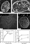
Role of FlnA expression in platelet morphology. (A and B) Representative cytoskeleton from a control FlnA+/y GATA1-Cre (A) or a FlnAloxP/y GATA1-Cre (B) resting platelet. Resting platelet cytoskeletons were prepared for the electron microscope by attachment to polylysine-coated coverslips in a permeabilizing PHEM buffer containing 0.75% Triton X-100, followed by fixation with 1% glutaraldehyde, and freezing in water, freeze drying, and metal casting. The FlnA-null cytoskeleton is enlarged and has a marginal microtubule ring, but the bulk of the cytoplasmic F-actin dissociates because of insufficient cross-linking of the actin filaments in the absence of FlnA. Bars, 0.2 µm. (C and D) Structure of the active cytoskeleton. FlnA+/y GATA1-Cre platelets (C) or FlnAloxP/y GATA1-Cre (D) were adhered to CRP-coated coverslips by centrifugation and incubated for 10 min at 37°C. Cells were then permeabilized in Triton X-100 in PHEM buffer containing 0.01% glutaraldehyde and processed for the electron microscopy as described. Bars, 0.5 µm. (Insets) Platelets were attached to and incubated on CRP-coated coverslips as in C and D and fixed with 3.7% formaldehyde in PBS for 30 min. F-actin was stained using 0.1% Triton X-100 in PBS and 0.1 µm Alexa Fluor 568 phalloidin. Bars, 5 µm. (E and F) Actin assembly. FlnA+/y GATA1-Cre and FlnAloxP/y GATA1-Cre platelets were activated with various concentrations of thrombin (E) or CRP (F) for 2 min at 37°C, as indicated. Platelets were fixed and permeabilized, washed, incubated with TRITC-labeled phalloidin, and analyzed by flow cytometry. Results are the ratio between the mean fluorescence of activated versus resting platelets and represent the mean ± SE of four independent experiments.
Figure 3.
Figure 3.
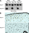
VWF receptor distribution in FlnA-null platelets. (A) GPIbα is not linked to the cytoskeleton of FlnA-null platelets. FlnA+/y GATA1-Cre and FlnAloxP/y GATA1-Cre platelets were activated or not (Rest) with 0.5 U/ml thrombin (Thr) or 3 µg/ml CRP for 5 min, as indicated. Triton X-100 soluble (Sup) and insoluble (Pel) fractions were collected by centrifugation of platelet lysates at 100,000 g for 30 min at 4°C, subjected to SDS-PAGE, and probed with a rat anti-GPIbα antibody. Results are representative of three independent experiments. (B) Distribution of anti-GPIbα gold on the surface of FlnA+/y GATA1-Cre and FlnAloxP/y GATA1-Cre platelets. GPIbα is found in linear arrays on the WT platelet surface. Arrays are highlighted with yellow shading. Particle counts revealed that 74.9 ± 5.9% of the total surface gold are in linear arrays composed of three or more particles. Only 13.1 ± 6.0% of the gold was arrayed on the FlnA-null platelet surface. Results are representative of five independent platelets for each mouse (0.5-µm2 area on each).
Figure 4.
Figure 4.
Platelet adhesion to collagen-bound VWF under arterial shear conditions. (A) After 3 min of perfusion of identical concentrations of labeled platelets in whole blood over collagen-coated surfaces at 1,500/s, ∼20% of the surface was covered by FlnA+/y GATA1-Cre platelets. Bar, 50 µm. (B) Mean of FlnA+/y GATA1-Cre platelets was set to 100% for better comparability. FlnAloxP/y GATA1-Cre platelet fluorescence covered 70% less area than FlnA+/y GATA1-Cre platelets. Error bars represent mean ± SEM (n = 6 mice). (C) Dwell time of 60 individual platelets per group under the same experimental conditions was significantly shorter in FlnAloxP/y than in FlnA+/y GATA1-Cre platelets. One-fifth of FlnA-null platelets detached after <500 ms under arterial shear conditions, whereas all FlnA+/y GATA1-Cre platelets dwelled for >500 ms. Results are representative of three independent experiments.
Figure 5.
Figure 5.
Functional defects in FlnA-null platelets. (A) FlnA+/y GATA1-Cre and FlnAloxP/y GATA1-Cre platelets in platelet-rich plasma (PRP) were activated with 10 µM ADP, 10 µM U46619, and/or 10 µM epinephrine (Epi), as indicated, and directly stained with PE-labeled JON/A for 10 min at room temperature. Platelets were then analyzed by flow cytometry. Results are the ratio between the mean fluorescence of activated versus resting platelets and represent the mean ± SD of four independent experiments. FlnA+/y GATA1-Cre and FlnAloxP/y GATA1-Cre platelets were activated with 50 nM PMA (B and G) or various concentrations of thrombin (C and H), CRP (D and I), convulxin (E and J), or rhodocytin (F and K) for 2 min at 37°C, as indicated. Platelets were then incubated with an FITC-labeled anti–mouse P-selectin antibody (B–F) or with Oregon green 488–labeled fibrinogen (G–K) and analyzed by flow cytometry. Results are expressed as percentage of positive platelets and represent mean ± SE of four independent experiments.
Figure 6.
Figure 6.
Protein tyrosine phosphorylation in platelets. FlnA+/y GATA1-Cre and FlnAloxP/y GATA1-Cre platelets were activated with CRP (A), convulxin (B), or rhodocytin (C) for 2 min at 37°C as indicated. Platelet lysates and Syk and PLC-γ2 were subjected to SDS-PAGE and probed with anti-Syk, anti-PLC-γ2, and anti-phosphotyrosine antibodies as indicated. Results are representative of three independent experiments.
Figure 7.
Figure 7.
Syk associates with FlnA Ig repeat 5. (A) FlnA and Syk associate in platelets. Human platelets were activated, or not, with 3 µg/ml CRP for 2 min at 37°C. Syk and FlnA were immunoprecipitated with rabbit antibodies N19 and 4762, respectively. Immunoprecipitates were subjected to SDS-PAGE and probed for FlnA. A control rabbit IgG was used as negative control for specificity. Results are representative of two experiments. (B) FlnA restricts Syk to the periphery of spread platelets. FlnA+/y GATA1-Cre and FlnAloxP/y GATA1-Cre platelets were attached by centrifugation at 280 g for 5 min on CRP-coated coverslips, incubated by 5 min at 37°C, fixed with 3.7% formaldehyde for 20 min, permeabilized with 0.1% Triton in 1% BSA/PBS, and incubated with anti-Syk antibody BR15 followed by a secondary Alexa Fluor 488–conjugated anti–rabbit Ig antibody in PBS/1% BSA containing 0.1 µM rhodamine-phalloidin. Bar, 5 µm. Results are representative of two experiments. (C) 100 nM His-tagged FlnA recombinant truncates containing Ig repeats 1–8+24, 8–16+24, or 17–24 were incubated with 25 nM GST-Syk or GST alone as control. Complexes were pulled down with glutathione Sepharose beads, subjected to SDS-PAGE, and probed for His (FlnA) as indicated. Results are representative of four experiments. (D) 50 nM eGFP-tagged FlnA repeat fragments containing full-length FlnA (FL), its actin-binding domain (ABD), or Ig repeats 1–3, 4, 5, 6–7, or 8 were incubated with 500 nM GST-Syk. Complexes were pulled down with glutathione Sepharose beads, subjected to SDS-PAGE, and probed for eGFP (FlnA). Results are representative of four experiments.

References

    1. Asazuma N., Ozaki Y., Satoh K., Yatomi Y., Handa M., Fujimura Y., Miura S., Kume S. 1997. Glycoprotein Ib-von Willebrand factor interactions activate tyrosine kinases in human platelets. Blood. 90:4789–4798 - PubMed
    1. Boylan B., Berndt M.C., Kahn M.L., Newman P.J. 2006. Activation-independent, antibody-mediated removal of GPVI from circulating human platelets: development of a novel NOD/SCID mouse model to evaluate the in vivo effectiveness of anti-human platelet agents. Blood. 108:908–914 10.1182/blood-2005-07-2937 - DOI - PMC - PubMed
    1. Boylan B., Gao C., Rathore V., Gill J.C., Newman D.K., Newman P.J. 2008. Identification of FcgammaRIIa as the ITAM-bearing receptor mediating alphaIIbbeta3 outside-in integrin signaling in human platelets. Blood. 112:2780–2786 10.1182/blood-2008-02-142125 - DOI - PMC - PubMed
    1. Clark E.A., Shattil S.J., Ginsberg M.H., Bolen J., Brugge J.S. 1994. Regulation of the protein tyrosine kinase pp72syk by platelet agonists and the integrin α IIb beta 3. J. Biol. Chem. 269:28859–28864 - PubMed
    1. Cranmer S.L., Ulsemer P., Cooke B.M., Salem H.H., de la Salle C., Lanza F., Jackson S.P. 1999. Glycoprotein (GP) Ib-IX-transfected cells roll on a von Willebrand factor matrix under flow. Importance of the GPib/actin-binding protein (ABP-280) interaction in maintaining adhesion under high shear. J. Biol. Chem. 274:6097–6106 10.1074/jbc.274.10.6097 - DOI - PubMed

Publication types

MeSH terms