. 2010 Aug 30;207(9):2019-31.
doi: 10.1084/jem.20090766. Epub 2010 Aug 16.

Viral entry and escape from antibody-mediated neutralization influence hepatitis C virus reinfection in liver transplantation

Affiliations

Viral entry and escape from antibody-mediated neutralization influence hepatitis C virus reinfection in liver transplantation

Samira Fafi-Kremer et al. J Exp Med. .

Abstract

End-stage liver disease caused by chronic hepatitis C virus (HCV) infection is a leading cause for liver transplantation (LT). Due to viral evasion from host immune responses and the absence of preventive antiviral strategies, reinfection of the graft is universal. The mechanisms by which the virus evades host immunity to reinfect the liver graft are unknown. In a longitudinal analysis of six HCV-infected patients undergoing LT, we demonstrate that HCV variants reinfecting the liver graft were characterized by efficient entry and poor neutralization by antibodies present in pretransplant serum compared with variants not detected after transplantation. Monoclonal antibodies directed against HCV envelope glycoproteins or a cellular entry factor efficiently cross-neutralized infection of human hepatocytes by patient-derived viral isolates that were resistant to autologous host-neutralizing responses. These findings provide significant insights into the molecular mechanisms of viral evasion during HCV reinfection and suggest that viral entry is a viable target for prevention of HCV reinfection of the liver graft.


Figures

Figure 1.
Figure 1.
Phylogenetic analyses of viral quasispecies evolution before and 7 d after LT. Evolution of HCV variants before and 7 d after LT. Rooted neighbor-joining trees of HCV HVR1 amino acid sequences from the six patients depicted in Table I are shown. Bootstrap values are expressed as percentages per 1,000 replicates. Only bootstrap proportions >70 are indicated. Sequences of variants isolated before LT and not detected on day 7 after transplantation are depicted as open black circles, sequences of variants isolated before LT and reinfecting the graft are depicted as open blue circles, and sequences isolated 7 d after LT are depicted as blue squares. Viral isolates used for functional analysis are indicated with capital letters.
Figure 2.
Figure 2.
Evolution of HCV variants before and 7 d after LT. Distribution of full-length E1E2 variants (mean number of clones per patient, 24; range, 20–31) depicted in Fig. 1 is shown for each patient. Circle graphs represent the percentage of each clone detected. The number of each clone is indicated. Viral isolates are indicated by an individual color and capital letters. Variants containing stop codons, insertions, or deletions altering the HCV open reading frame are depicted with a number sign (#) and were not further analyzed in HCVpp assays. Variants reinfecting the liver graft are depicted in blue, and nonselected variants not detected after transplantation are depicted in white, gray, or black. BT, before LT; D7, 7 d after LT
Figure 3.
Figure 3.
Viral entry is important for selection of viral variants after LT. (A) Transfection efficiency was analyzed for each variant and control (Ctrl; empty vector without E1E2) by quantifying luciferase expression (expressed as normalized percentage of transfection efficiency based on the predominant selected variant). (B) Comparative analysis of viral entry of HCVpps containing full-length E1E2 functional envelope proteins from pretransplant variants in Huh7 cells depicted in Fig. 2. HCVpps bearing patient-derived HCV envelope glycoproteins were added to Huh7 cells, and infection was analyzed by luciferase reporter gene expression. Results are expressed in relative light units (RLU) plotted in a logarithmic scale. The threshold for a detectable infection in this system is indicated by dashed lines. The detection limit for positive luciferase reporter protein expression was 3 × 10³ RLU/assay, corresponding to the mean ± 3 SD of background levels, i.e., luciferase activity of naive noninfected cells or cells infected with pseudotypes without HCV envelopes (Dimitrova et al., 2008). Background levels of the assay were determined in each experiment. Means ± SD from at least four independent experiments (performed in triplicate) are shown. Statistically significant differences (repeated measures ANOVA) in HCVpp entry between the predominant selected strain and nonselected strains are indicated by asterisks (**, P < 0.001). Variants reinfecting the liver graft are depicted in blue, and nonselected variants not detected after transplantation are depicted in white, gray, or black.
Figure 4.
Figure 4.
Escape from antibody-mediated neutralization is a key determinant for selection of viral variants after LT. HCVpps were incubated with pretransplant anti-HCV–positive serum or control anti-HCV–negative serum (Ctrl) in serial dilutions for 1 h at 37°C. HCVpp–antibody complexes were then added to Huh7 cells, and infection was analyzed as described in Fig. 3. Calculation of neutralization and determination of background and thresholds for neutralization are described in Materials and methods. End point dilution titers are indicated for each variant. Dashed lines indicate the threshold for a positive neutralization titer corresponding to 1:40. Variants reinfecting the liver graft are depicted in blue, and nonselected variants not detected after transplantation are depicted in white, gray, or black. Means from at least four independent experiments (performed in triplicate) are shown. Statistically significant differences (repeated measures ANOVA) in neutralization between the predominant selected strain and nonselected strains are indicated by asterisks (**, P < 0.001). NI, noninfectious.
Figure 5.
Figure 5.
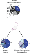
Evolution of HCV variants in the uPA-SCID mouse model infected with patient-derived pretransplant serum. A human liver–chimeric mouse was challenged by intraperitoneal injection of pretransplant serum of patient P01 (infectious dose administered, 3.3 × 10⁵ IU) and sacrificed at week 3 after infection. HCV RNA was isolated and amplified by RT-PCR from liver and plasma as described previously (Schvoerer et al., 2007). HCV isolates in plasma and liver samples were cloned and sequenced as described in Materials and methods (30 clones per sample in one animal). Circle graphs represent the percentage of each clone detected, and numbers of clones are shown. Variant VL, which was selected after LT in patient P01, was the most prevalent variant in both plasma and liver of the infected uPA-SCID mouse. Variants containing stop codons, insertions, or deletions altering the HCV open reading frame are depicted with a number sign (#).
Figure 6.
Figure 6.
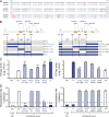
Mutations in envelope region E2425–483 mediate enhanced viral entry and escape from neutralizing antibodies. To map envelope regions mediating enhanced entry and viral escape, we exchanged four regions spanning C-terminal E1 and N-terminal E2, E1, E2- HVR1, and E2-HVR2 of the envelope glycoproteins of selected variant VL and nonselected variant VC of patient P01 (see Figs. 2–4). These regions include aa 221–483, aa 221–357, aa 358–424, and aa 425–483, respectively. (A) Deduced amino acid sequences of the exchanged region between P01VC (black) and P01VL (blue). Amino acid changes are indicated in red bold letters. (B) Construction of recombinant chimeric HCVpps P01VCVL221–483 and P01VLVC221–483, P01VCVL-E1 and P01VLVC-E1, P01VCVL-HVR1 and P01VLVC-HVR1, and P01VCVL-HVR2 and P01VLVC-HVR2 by exchanging E1E2 envelope domain aa 221–483, aa 221–357, aa 358–424, and aa 425–483, respectively, of nonselected variant VC (patient 01) depicted in white and escape isolate VL (patient 01) depicted in blue (see Figs. 2–4). HVR1 and HVR2 are shown in orange, and CD81 binding domains (CD81 BD) are shown in green. Positions of E2 epitopes I and II are indicated (Zhang et al., 2007, 2009). The number of mutations within each region is shown. (C) Viral entry of HCVpps containing chimeric envelope proteins in Huh7 cells. HCVpps were incubated with Huh7 cells, and infection was analyzed as described in Fig. 3. Results are expressed in relative light units (RLU) plotted in a logarithmic scale. The threshold for a detectable infection is 3 × 10³ RLU and was determined as described in Fig. 3. (D) Neutralization of HCVpps by autologous pretransplant serum was performed as described in Fig. 4. End point dilution titers are indicated for each variant. Dashed lines indicate the threshold for a positive neutralization titer corresponding to 1:40. Calculation of neutralization and determination of threshold titers are described in Materials and methods. Chimeric HCVpps are depicted in dashed blue. Statistically significant differences (repeated measures ANOVA) in HCVpp entry or neutralization between VC and VL wild-type and mutant variants are indicated by asterisks (**, P < 0.001). Ctrl, negative control; MT, mutation.
Figure 7.
Figure 7.
Cross-neutralization of escape variants infecting the liver graft by antienvelope and antireceptor mAbs. (A) Neutralization of HCVpps from viral isolates by cross-neutralizing anti-E2 mAb AP33. HCVpps derived from different isolates were incubated with 10 µg/ml anti-E2 AP33 or isotype monoclonal control IgG, and neutralization of viral entry in Huh7 cells was determined as described in Fig. 4 (entry in the presence of isotype monoclonal control IgG = 100%). Neutralization was calculated as described in Materials and methods. Means ± SD from three independent experiments (performed in triplicate) are shown. (B and C) Neutralization of HCV isolates having escaped patients’ neutralizing responses by anti-E2 and anti-CD81 mAbs in primary human hepatocytes. HCVpps derived from viral isolates selected after LT (P01VL, P02VI, P03VC, P04VE, P05VF, and P06VI) were incubated with serial dilutions of anti-E2 AP33 (B) or control IgG from mouse (Ctrl) as described in Fig. 4 and then added to primary human hepatocytes. For analysis of neutralization using anti-CD81, primary human hepatocytes were preincubated with anti-CD81 or isotype control IgG for 1 h at 37° before incubation with patient-derived HCVpps (C). Means from one representative experiment (performed in triplicate) out of two independent experiments are shown. 50% neutralization of HCVpp entry is indicated by a dashed line.

Comment in

Similar articles

Cited by

References

    1. Aurora R., Donlin M.J., Cannon N.A., Tavis J.E. 2009. Genome-wide hepatitis C virus amino acid covariance networks can predict response to antiviral therapy in humans. J. Clin. Invest. 119:225–236 - PMC - PubMed
    1. Barth H., Schnober E.K., Zhang F., Linhardt R.J., Depla E., Boson B., Cosset F.L., Patel A.H., Blum H.E., Baumert T.F. 2006. Viral and cellular determinants of the hepatitis C virus envelope-heparan sulfate interaction. J. Virol. 80:10579–10590 10.1128/JVI.00941-06 - DOI - PMC - PubMed
    1. Bartosch B., Dubuisson J., Cosset F.L. 2003. Infectious hepatitis C virus pseudo-particles containing functional E1–E2 envelope protein complexes. J. Exp. Med. 197:633–642 10.1084/jem.20021756 - DOI - PMC - PubMed
    1. Bartosch B., Verney G., Dreux M., Donot P., Morice Y., Penin F., Pawlotsky J.M., Lavillette D., Cosset F.L. 2005. An interplay between hypervariable region 1 of the hepatitis C virus E2 glycoprotein, the scavenger receptor BI, and high-density lipoprotein promotes both enhancement of infection and protection against neutralizing antibodies. J. Virol. 79:8217–8229 10.1128/JVI.79.13.8217-8229.2005 - DOI - PMC - PubMed
    1. Bowen D.G., Walker C.M. 2005. Adaptive immune responses in acute and chronic hepatitis C virus infection. Nature. 436:946–952 10.1038/nature04079 - DOI - PubMed

MeSH terms