. 2010 Aug 23;190(4):637-50.
doi: 10.1083/jcb.200908092. Epub 2010 Aug 16.

BAG-6 is essential for selective elimination of defective proteasomal substrates

Affiliations

BAG-6 is essential for selective elimination of defective proteasomal substrates

Ryosuke Minami et al. J Cell Biol. .

Abstract

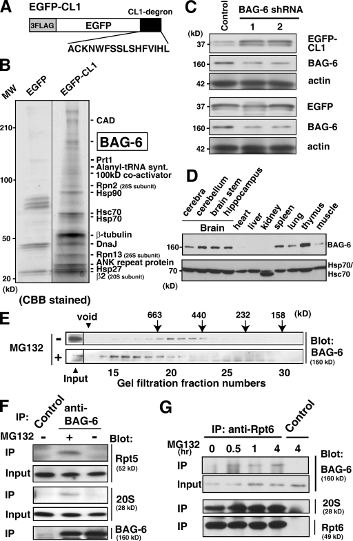
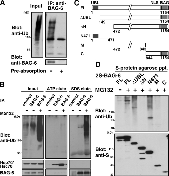
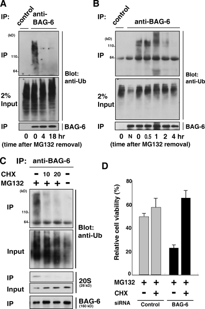
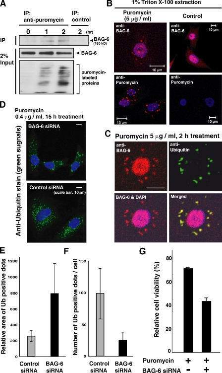
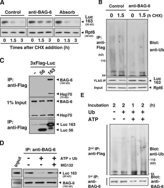
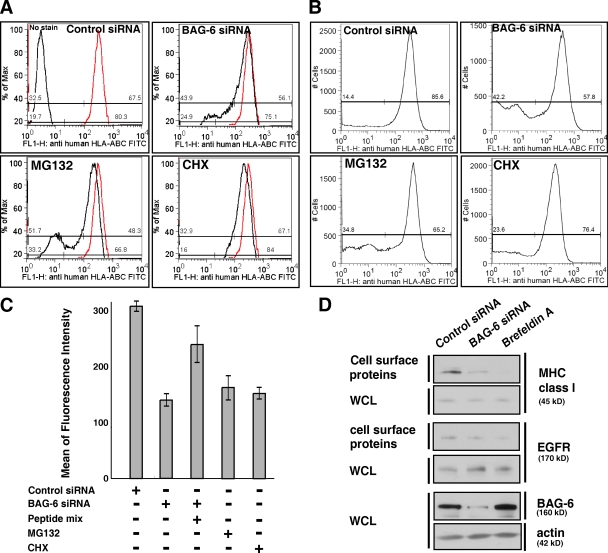
BAG-6/Scythe/BAT3 is a ubiquitin-like protein that was originally reported to be the product of a novel gene located within the human major histocompatibility complex, although the mechanisms of its function remain largely obscure. Here, we demonstrate the involvement of BAG-6 in the degradation of a CL1 model defective protein substrate in mammalian cells. We show that BAG-6 is essential for not only model substrate degradation but also the ubiquitin-mediated metabolism of newly synthesized defective polypeptides. Furthermore, our in vivo and in vitro analysis shows that BAG-6 interacts physically with puromycin-labeled nascent chain polypeptides and regulates their proteasome-mediated degradation. Finally, we show that knockdown of BAG-6 results in the suppressed presentation of MHC class I on the cell surface, a procedure known to be affected by the efficiency of metabolism of defective ribosomal products. Therefore, we propose that BAG-6 is necessary for ubiquitin-mediated degradation of newly synthesized defective polypeptides.


Figures

Figure 1.
Figure 1.
BAG-6 is essential for CL1 degron-dependent proteasomal degradation. (A) Schematic representation of the 3xFlag-tagged EGFP protein fused with CL1 degron used in this study. (B) CL1 degron-associated proteins identified in this study. After transfection of a 3xFlag-tagged EGFP-CL1 expression vector, HeLa cells were treated with 5 µM MG132 for 4.5 h. Proteins immunoprecipitated with antibody against Flag were subjected to SDS-PAGE and PMF analysis. 3xFlag-tagged EGFP immunoprecipitates were used as a negative control. (C) Knockdown of BAG-6 suppressed the degradation of the CL1 degron substrate. 3xFlag-tagged EGFP-CL1 was expressed in HeLa cells with two distinct shRNA vectors for BAG-6 (siRNA-1 and siRNA-2) or control siRNA. After 60 h of shRNA treatment, whole-cell extracts were prepared and subjected to immunoblot analysis with antibodies against Flag, BAG-6, and actin. (D) Expression patterns of endogenous BAG-6 protein in various adult mouse tissues (top). The anti-Hsp70/Hsc70 blot confirmed equal protein loading (bottom). (E) MG132 treatment stimulated the formation of a larger BAG-6 complex. Extracts of NIH3T3 cells were subjected to gel filtration with Superose 6, and the fractions were subjected to Western blotting with specific antibodies against BAG-6. Cells were cultured with (+) or without (−) 20 µM MG132 before harvesting. (F and G) BAG-6 was associated with the 26S proteasome in HeLa cells. (F) An antibody against BAG-6 was used to immunoprecipitate endogenous BAG-6 protein from extracts of HeLa cells cultured with (+) or without (−) 20 µM MG132 before harvesting. The precipitates were immunoblotted with antibodies to the 19S complex (Rpt5 subunit), 20S proteasome (α5 subunit), and BAG-6. (G) Using an antibody against the 26S proteasome subunit Rpt6, endogenous 26S proteasomes were immunoprecipitated from HeLa cell extracts. HeLa cells were treated with 20 µM MG132 for indicated periods. Immunoglobulin derived from nonimmune mouse serum was used as a negative control. The precipitates were immunoblotted with antibodies against BAG-6, 20S, and 19S complex (Rpt6 subunit).
Figure 2.
Figure 2.
BAG-6 associates with polyubiquitinated proteasomal substrates. (A) Endogenous BAG-6 protein was affinity purified from extracts of MG132-treated HeLa cells with an antibody against BAG-6, and the precipitates were immunoblotted with antibodies against ubiquitin and BAG-6. Ubiquitin coprecipitation of BAG-6 was abolished by preabsorption with an excess of BAG-6 antigen. (B) Endogenous BAG-6 protein precipitated from HeLa cell extracts was treated with 5 mM ATP (ATP-elute) before boiling in 1% SDS (SDS-elute). Each eluate was immunoblotted with antibodies against ubiquitin, Hsp70/Hsc70, and BAG-6. Cells were treated with (+) or without (−) 20 µM MG132 for 6 h before being harvested as indicated. Immunoglobulin derived from nonimmune rabbit serum was used in immunoprecipitation as a negative control. (C) Schematic representation of the BAG-6 deletion mutant proteins used in this study. The numbers denote corresponding amino acid numbers. (D) N-terminal 471 amino acids of BAG-6 were essential for the binding with polyubiquitinated proteins. The full-length (FL) form of 2S-tagged BAG-6 and its truncated derivatives were expressed in HeLa cells as indicated. Before harvesting, cells were treated with or without 10 µM MG132 for 12 h. Each form of BAG-6 was affinity purified with S-protein agarose, and the bound materials were blotted with antibodies against ubiquitin and S peptide.
Figure 3.
Figure 3.
BAG-6 associates with polyubiquitinated newly synthesized defective proteins. (A and B) Polyubiquitinated proteins associated with BAG-6 were degraded in vivo. HeLa cells were treated with MG132 (5 µM) for 4.5 h, the inhibitor was then washed out (this time point being defined as time zero), and the cells were cultured in a fresh medium (without MG132) for the indicated times. After harvesting the cells, endogenous BAG-6 was immunoprecipitated and the coprecipitated materials were probed with an antibody against ubiquitin. Normal rabbit immunoglobulin was used as a negative control for the immunoprecipitation procedures. “N” indicates no addition of MG132 to the cell culture. (C) Polyubiquitinated proteins associated with BAG-6 were abolished by addition of the translation inhibitor cycloheximide (CHX). HeLa cells were treated with 5 µM MG132 with or without 10 µg/ml or 20 µg/ml CHX as indicated for 1 h and then subjected to immunoprecipitation using antibody against BAG-6. The immunoprecipitates were probed with antibodies against ubiquitin, 20S proteasome, and BAG-6. (D) Depletion of BAG-6 made HeLa cells more sensitive to proteasome inhibitor-induced cell death, but the effect of BAG-6 knockdown was abrogated by adding CHX. 48 h after treatment with siRNA, cells were treated with 5 µM MG132 for 24 h with or without 10 µg/ml CHX, and cell viability was analyzed using an MTT cell counting system. The percent viability was normalized to the cells transfected with control siRNA without MG132 treatment (arbitrarily assigned a value of 100%). Error bars represent SD calculated from three experiments. P < 0.01, by t test.
Figure 4.
Figure 4.
BAG-6 is involved in the metabolism of puromycin-induced nascent chain polypeptides. (A) BAG-6 bound to truncated puromycin-labeled proteins. After treatment with 5 µg/ml puromycin for the indicated times, HeLa cells were harvested, the puromycin-labeled polypeptides were immunoprecipitated from cell extracts with antibody against puromycin, and the precipitates were probed with antibody against BAG-6. (B and C) BAG-6 localized on cytoplasmic dots after puromycin treatment. HeLa cells were treated with 5 µg/ml puromycin for 2 h and extracted with 1% Triton X-100. Fixed cells were stained with antibodies against BAG-6, puromycin (B), and ubiquitin (C). Note that a part of BAG-6 also localized in the nucleus. (D–F) BAG-6 controlled the dynamics of puromycin-induced aggregate formation. Puromycin-treated HeLa cells with BAG-6 siRNA contained enlarged but reduced numbers of ubiquitin-positive cytoplasmic aggregates. Error bars represent SD calculated from three experiments. (G) 48 h after siRNA treatment, cells were treated with puromycin for 24 h and cell viability was analyzed with a cell counting kit. The percent viability was normalized to the cells without puromycin treatment.
Figure 5.
Figure 5.
BAG-6 provides a platform that is necessary for linking puromycin-labeled defective protein with degradation machinery. (A) Addition of antibody against BAG-6 inhibited the degradation of puromycin-labeled, truncated luciferase in vitro. A messenger RNA encoding the 3xFlag-tagged N-terminal 163 residues of luciferase (Luc163) was incubated in a rabbit reticulocyte lysate chasing with 2 mM puromycin addition. After addition of 50 µg/ml anti–BAG-6 antibody, cycloheximide (CHX) chase analysis was performed, and lysates were harvested at the indicated times and probed with antibodies against Flag and Rpt6 to evaluate the stability of puromycin-labeled Luc163 (Luc163). Non-immune rabbit IgG or antibody against BAG-6 that was preabsorbed with an excess amount of recombinant antigen was used as a negative control. (B) Addition of antibody against BAG-6 inhibited ubiquitination of puromycin-labeled, truncated luciferase. Blot with Flag-Luc163 and Rpt6 are indicated as loading controls. (C) Immunoprecipitation of in vitro translated Luc163 with anti-Flag M2 beads coprecipitated endogenous BAG-6 and Hsp70 from lysates. (D) BAG-6 provided a platform for the targeted degradation of Luc163. Endogenous BAG-6 was immunoprecipitated from a rabbit reticulocyte lysate that was translating 3xFlag-tagged Luc163. The precipitated immunocomplex was further incubated in the presence (+) or absence (−) of 5 mM ATP, ubiquitin, and 25 µM MG132 at 37°C for 2 h as indicated. After incubation, the precipitated complexes were subjected to Western blot analysis with an antibody against Flag to examine the stability of Luc163 on BAG-6 during the incubation periods. (E) Endogenous BAG-6 was immunoprecipitated (first IP) as in D, and the precipitated complex was incubated with MG132 under the conditions indicated. After incubation, the complexes were denatured by SDS and diluted samples were further subjected to precipitation with anti-Flag M2 agarose (second IP). Precipitated, Flag-tagged Luc163 was blotted with antibody against ubiquitin to estimate the extent of its modification with ubiquitin.
Figure 6.
Figure 6.
BAG-6 modulates expression of MHC class I molecules on the cell surface. (A and B) Live-cell flow cytometric analysis of HeLa cells with FITC-labeled antibody against MHC class I that recognizes cell surface HLA-ABC antigen. As positive controls, the results of treatment with 10 µM MG132 (A and B) and 10 µg/ml CHX (A) are shown. The data were obtained by linear scale analysis (A) and by log scale analysis (B). The flow cytometric pattern of negative control siRNA is indicated as a red line, and those after treatment with BAG-6 siRNA, MG132, and CHX are indicated with black lines (A). BAG-6 knockdown was performed with two independent duplex siRNAs as described in Materials and methods, and both siRNA gave a similar result. Representatives are results with duplex siRNAs of BAG-6-2 (5′-ATGATGCACATGAACATTC-3′). (C) Quantitative evaluations of the mean of fluorescence intensity of FITC-HLA-ABC on the surface of HeLa cells. Antigenic peptide mixture was pulsed with cells at 50 µM for 16 h. The data shown are the results of at least three independent experiments. (D) Biotin-labeling experiments. Cell surface proteins were biotinylated and affinity purified with avidin beads and blotted with antibody against MHC class I. Knockdown of BAG-6 reduced the expression of MHC class I on the cell surface, whereas the amount of EGF receptor (EGFR) on the cell surface was not affected. Brefeldin A treatment was used as a positive control for general suppression of transport of MHC class I and EGFR to the cell surface. Actin blots for whole-cell lysates (WCL) were used as a loading control for whole cellular proteins.

Similar articles

Cited by

References

    1. Anton L.C., Yewdell J.W., Bennink J.R. 1997. MHC class I-associated peptides produced from endogenous gene products with vastly different efficiencies. J. Immunol. 158:2535–2542 - PubMed
    1. Ardley H.C., Scott G.B., Rose S.A., Tan N.G., Markham A.F., Robinson P.A. 2003. Inhibition of proteasomal activity causes inclusion formation in neuronal and non-neuronal cells overexpressing Parkin. Mol. Biol. Cell. 14:4541–4556 10.1091/mbc.E03-02-0078 - DOI - PMC - PubMed
    1. Banerji J., Sands J., Strominger J.L., Spies T. 1990. A gene pair from the human major histocompatibility complex encodes large proline-rich proteins with multiple repeated motifs and a single ubiquitin-like domain. Proc. Natl. Acad. Sci. USA. 87:2374–2378 10.1073/pnas.87.6.2374 - DOI - PMC - PubMed
    1. Bence N.F., Sampat R.M., Kopito R.R. 2001. Impairment of the ubiquitin-proteasome system by protein aggregation. Science. 292:1552–1555 10.1126/science.292.5521.1552 - DOI - PubMed
    1. Chen L., Madura K. 2002. Rad23 promotes the targeting of proteolytic substrates to the proteasome. Mol. Cell. Biol. 22:4902–4913 10.1128/MCB.22.13.4902-4913.2002 - DOI - PMC - PubMed

Publication types

MeSH terms