. 2010 Aug 23;190(4):523-31.
doi: 10.1083/jcb.201002035. Epub 2010 Aug 16.

Autophagic degradation of dBruce controls DNA fragmentation in nurse cells during late Drosophila melanogaster oogenesis

Affiliations

Autophagic degradation of dBruce controls DNA fragmentation in nurse cells during late Drosophila melanogaster oogenesis

Ioannis P Nezis et al. J Cell Biol. .

Abstract

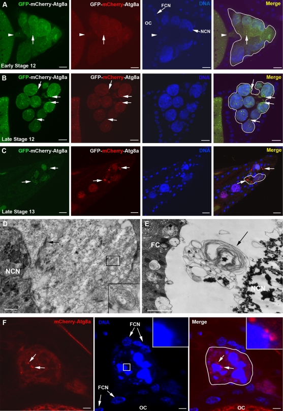
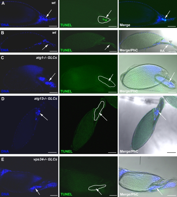
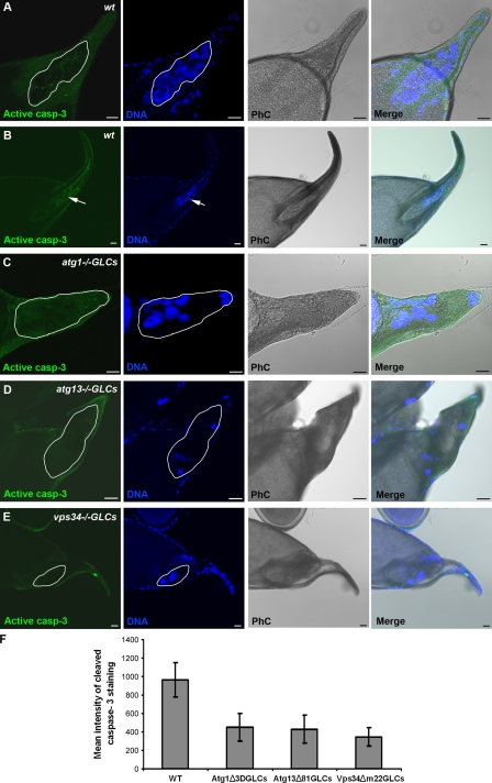
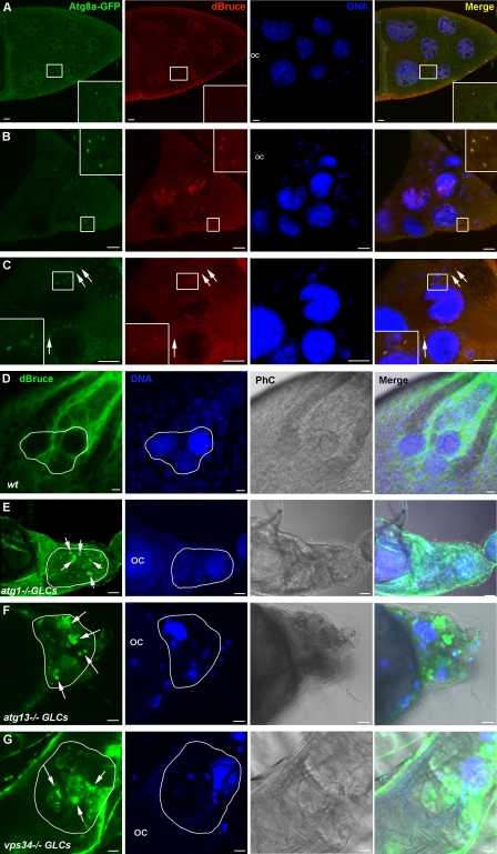
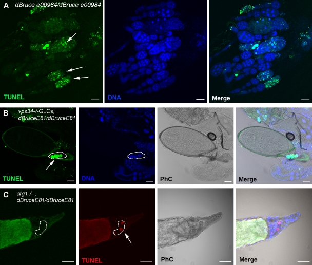
Autophagy is an evolutionarily conserved pathway responsible for degradation of cytoplasmic material via the lysosome. Although autophagy has been reported to contribute to cell death, the underlying mechanisms remain largely unknown. In this study, we show that autophagy controls DNA fragmentation during late oogenesis in Drosophila melanogaster. Inhibition of autophagy by genetically removing the function of the autophagy genes atg1, atg13, and vps34 resulted in late stage egg chambers that contained persisting nurse cell nuclei without fragmented DNA and attenuation of caspase-3 cleavage. The Drosophila inhibitor of apoptosis (IAP) dBruce was found to colocalize with the autophagic marker GFP-Atg8a and accumulated in autophagy mutants. Nurse cells lacking Atg1 or Vps34 in addition to dBruce contained persisting nurse cell nuclei with fragmented DNA. This indicates that autophagic degradation of dBruce controls DNA fragmentation in nurse cells. Our results reveal autophagic degradation of an IAP as a novel mechanism of triggering cell death and thereby provide a mechanistic link between autophagy and cell death.


Figures

Figure 1.
Figure 1.
Autophagy is activated in dying nurse cells during late oogenesis in D. melanogaster. (A–C) Confocal micrographs of egg chambers of flies expressing the UASp-GFP-mCherry-DrAtg8a transgene exclusively in the germline (genotype: UASp-GFP-mCherry-DrAtg8a/+; nanos-VP-16 Gal4/+). Expression of UASp-GFP-mCherry-DrAtg8a exhibits punctate staining pattern (arrows). Note that during developmental stages early 12 (A) and late 12 (B), the punctate dots are yellow (merge). In late stage 13 (C), large red dots are evident, indicating the presence of acidic compartments (autolysosomes; arrows). The arrowheads in A point to the physical connection between the oocyte and the nurse cells during early stage 12 (not observed during late stage 12). (D and E) Transmission electron micrographs of nurse cells in stage 12 (D) and in stage 13 (E) egg chambers. Autophagosomes (inset in D) and autolysosomes (arrow in E) are evident in the remaining nurse cell cytoplasm (arrow in D indicates actin bundles). Note that the autolysosome shown in E contains condensed material resembling the material of the condensed and fragmented nurse cell nucleus (NCN), providing evidence for nuclear autophagy. (F) Nurse cell of a late stage 13 egg chamber expressing UASp-mCherry-DrAtg8a exclusively in the germline (genotype: UASp-mCherry-DrAtg8a/+; nanos-VP-16 Gal4/+). mCherry-DrAtg8a puncta are attached to or located adjacent to the fragmented nucleus (arrows and insets). Hoechst staining (blue) was performed to visualize the nuclei. Nurse cells are outlined with a white line. FC, follicle cell; FCN, follicle cell nucleus; OC, oocyte. Bars: (A–C) 10 µm; (D–F) 1 µm.
Figure 2.
Figure 2.
Genetic inhibition of autophagy in the germline prevents DNA fragmentation. Confocal micrographs of stage 13/14 egg chambers after TUNEL staining. (A) Wild-type (wt) stage 13 egg chamber contains nurse cell nuclei exhibiting fragmented DNA (positive TUNEL staining; arrows). (B) Wild-type stage 14 egg chambers do not contain nurse cells. Arrows point to the respiratory appendages (RA). (C–E) Stage 14 atg1−/− (C), atg13−/− (D), and vps34−/− (E) germline mutant chambers have persisting nurse cell nuclei that do not contain fragmented DNA (negative TUNEL staining; arrows). Nurse cells are outlined with a white line. Draq5/Hoechst staining (blue) was performed to visualize the nuclei. PhC, phase contrast. Bars, 50 µm.
Figure 3.
Figure 3.
Germline autophagy mutant egg chambers exhibit reduced expression of cleaved caspase-3. Confocal micrographs of stage 13/14 egg chambers stained for cleaved caspase-3 (casp-3). (A) Wild-type (wt) late stage 13 egg chamber showing cleaved caspase-3 staining in the degenerating nurse cell cluster. (B) In wild-type stage 14 egg chamber, nurse cells are completely degraded (arrows). (C–E) atg1−/− (C), atg13−/− (D), and vps34−/− (E) germline mutant stage 14 egg chambers exhibit significantly reduced staining for cleaved caspase-3 and contain persisting nurse cell nuclei. Nurse cells are outlined with a white line. Draq5/Hoechst staining (blue) was performed to visualize the nuclei. PhC, phase contrast. (F) Quantification of mean intensity of cleaved caspase-3 staining observed in the nurse cells of stage 12–14 egg chambers in wild-type and germline autophagy mutant egg chambers. Wild type (WT): three independent experiments, n = 30 egg chambers; atg1−/− GLCs: four independent experiments, n = 30 egg chambers; atg13−/− GLCs: four independent experiments, n = 30 egg chambers; vps34−/− GLCs: three independent experiments, n = 30 egg chambers. Data are presented as mean ± SD. Difference was significant with P < 0.001 for all values versus wild type. Bars, 20 µm.
Figure 4.
Figure 4.
dBruce colocalizes with the autophagic marker Atg8a-GFP in the nurse cells during late oogenesis and is accumulated in autophagy germline mutants. Confocal micrographs of late stage egg chambers expressing Atg8a-GFP and stained for dBruce. (A–C) Stage 10 (A), early stage 12 (B), and late stage 12 (C). dBruce colocalizes with Atg8a-GFP in punctuate structures during stages 12 and 13 (insets in B and insets and arrows in C) and not during stage 10 (insets in A). (D–G) Confocal micrographs of stage 14 wild-type (D) and autophagy mutant egg chambers (E–G) stained for dBruce. dBruce is accumulated in atg1, atg13, and vps34 mutant nurse cells (arrows). Draq5/Hoechst staining (blue) was performed to visualize the nuclei. Nurse cells are outlined with a white line. OC, oocyte; PhC, phase contrast. Bars, 10 µm.
Figure 5.
Figure 5.
dBruce is required for nurse cell survival by controlling DNA fragmentation during oogenesis. (A) Confocal micrographs of dBrucee00984 mutant ovarioles stained for TUNEL and DNA. Arrows point to degenerating stage 8/9 egg chambers that are TUNEL positive. (B and C) Confocal micrographs of vps34/dBruceE81 and atg, dBruceE81 double mutant egg chambers stained for TUNEL (green and red, respectively) and DNA. (B) vps34−/−GLCs; dBruceE81/dBruceE81 stage 14 mutant egg chamber contain persisting TUNEL-positive nurse cell nuclei (outlined; arrow). (C) atg1−/−GLCs, dBruceE81/dBruceE81 stage 14 mutant egg chamber contain persisting TUNEL-positive nurse cell nuclei. atg1 mutant nurse cells are identified by the lack of GFP staining (outlined; arrow). Draq5/Hoechst staining (blue) was performed to visualize the nuclei. PhC, phase contrast. Bars, 50 µm.

References

    1. Arama E., Agapite J., Steller H. 2003. Caspase activity and a specific cytochrome C are required for sperm differentiation in Drosophila. Dev. Cell. 4:687–697 10.1016/S1534-5807(03)00120-5 - DOI - PubMed
    1. Arama E., Bader M., Rieckhof G.E., Steller H. 2007. A ubiquitin ligase complex regulates caspase activation during sperm differentiation in Drosophila. PLoS Biol. 5:e251 10.1371/journal.pbio.0050251 - DOI - PMC - PubMed
    1. Bass B.P., Tanner E.A., Mateos San Martín D., Blute T., Kinser R.D., Dolph P.J., McCall K. 2009. Cell-autonomous requirement for DNaseII in nonapoptotic cell death. Cell Death Differ. 16:1362–1371 10.1038/cdd.2009.79 - DOI - PMC - PubMed
    1. Berry D.L., Baehrecke E.H. 2007. Growth arrest and autophagy are required for salivary gland cell degradation in Drosophila. Cell. 131:1137–1148 10.1016/j.cell.2007.10.048 - DOI - PMC - PubMed
    1. Cavaliere V., Taddei C., Gargiulo G. 1998. Apoptosis of nurse cells at the late stages of oogenesis of Drosophila melanogaster. Dev. Genes Evol. 208:106–112 10.1007/s004270050160 - DOI - PubMed

Publication types

MeSH terms