. 2010 Nov;78(11):4691-6.
doi: 10.1128/IAI.00801-10. Epub 2010 Aug 16.

Escherichia coli subtilase cytotoxin induces apoptosis regulated by host Bcl-2 family proteins Bax/Bak

Affiliations

Escherichia coli subtilase cytotoxin induces apoptosis regulated by host Bcl-2 family proteins Bax/Bak

Kerrie L May et al. Infect Immun. 2010 Nov.

Abstract

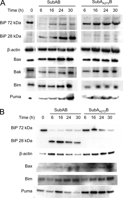
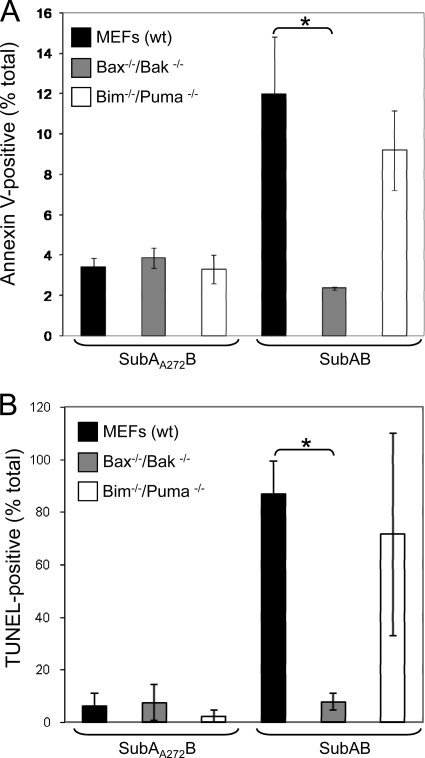
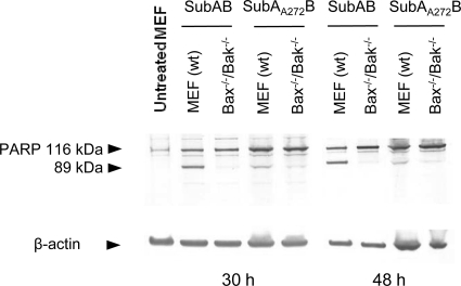
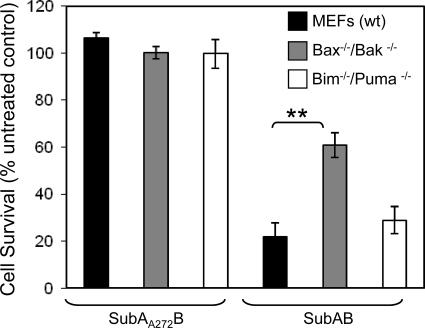
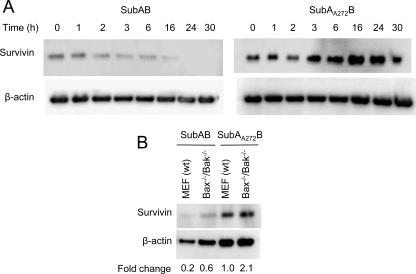
Subtilase cytotoxin (SubAB) was first isolated from a Shiga toxigenic Escherichia coli (STEC) strain that was responsible for an outbreak of hemolytic-uremic syndrome and is the prototype of a new family of AB(5) cytotoxins. SubAB is a subtilase-like serine protease, and upon uptake by host cells, it is trafficked to the endoplasmic reticulum (ER), where it cleaves the essential ER chaperone BiP (GRP78) with high specificity. Previous work has shown that BiP cleavage by SubAB initiates ER stress-signaling pathways in host cells that eventuate in cell death associated with DNA fragmentation, a hallmark of apoptosis. The present study has investigated the role of the Bcl-2 protein family, which has been shown to regulate ER stress-induced apoptosis in other model systems. Examination of the cytotoxicity of SubAB for wild-type and bax(-/-)/bak(-/-) mouse embryonic fibroblasts and comparison of apoptotic markers in these cells revealed that SubAB cytotoxicity can be predominantly attributed to the activation of apoptotic pathways activated by Bax/Bak. The results of the present study further our understanding of the molecular mechanism whereby SubAB kills eukaryotic cells and contributes to STEC pathogenesis, in addition to consolidating the roles of Bcl-2 family members in the regulation of ER stress-induced apoptosis.


Figures

FIG. 1.
FIG. 1.
Effect of SubAB on the levels of Bcl-2 protein family members. Vero cells (A) or MEFs (B) were treated with either SubAB (100 ng/ml) or SubAA272B (100 ng/ml) for the indicated times, and lysates were analyzed by Western blotting. β-Actin was used as an internal loading control, and BiP cleavage was used as a control for toxin activity. Data are representative of those from two independent experiments.
FIG. 2.
FIG. 2.
Quantitative analysis of SubAB-induced apoptosis in wt, bax−/−/bak−/−, and bim−/−/puma−/− MEFs. (A) In situ labeling of phosphatidylserine on the surface of apoptotic cells. Cells were treated with either SubAB (100 ng/ml) or SubAA272B (100 ng/ml) for 24 h. Phosphatidylserine was labeled with annexin V conjugated to Alexa 488 dye. (B) In situ TUNEL of DNA fragmentation in MEFs that were treated with either SubAB (100 ng/ml) or SubAA272B (100 ng/ml) for 30 h. Data represent the means ± SDs from two independent experiments. *, P < 0.05, Student's unpaired, two-tailed t test.
FIG. 3.
FIG. 3.
PARP processing in wt and bax−/−/bak−/− MEFs treated with SubAB. Cells were treated with either SubAB (100 ng/ml) or SubAA272B (100 ng/ml) for 30 h or 48 h, and lysates were analyzed by Western blotting with anti-PARP antibodies. β-Actin was used as an internal loading control. The mobilities of intact (116-kDa) and cleaved (89-kDa) PARP are indicated. Data are representative of those from two independent experiments.
FIG. 4.
FIG. 4.
Relative cytotoxicity of SubAB for wt, bax−/−/bak−/−, and bim−/−/puma−/− MEFs. Cells were treated with either SubAB (100 ng/ml) or SubAA272B (100 ng/ml) for 48 h, and cytotoxicity was measured using crystal violet staining. Data are expressed as percent cell survival compared to the rate of survival for untreated control cells. Data represent the means ± standard errors of the means from four independent experiments, **, P < 0.01, Student's unpaired, two-tailed t test.
FIG. 5.
FIG. 5.
Effect of SubAB on survivin protein levels. (A) Vero cells were treated with either SubAB (100 ng/ml) or SubAA272B (100 ng/ml) for 0 to 30 h, and lysates were analyzed by Western blotting with the appropriate antisurvivin antibody (see Materials and Methods). β-Actin was used as an internal loading control. (B) wt and bax−/−/bak−/− MEFs were treated with either SubAB (100 ng/ml) or SubAA272B (100 ng/ml) for 48 h, and lysates were analyzed by Western blotting with the appropriate antisurvivin antibody. β-Actin was used as an internal loading control. The fold change in survivin levels relative to the level in SubAA272B-treated wt MEFs was calculated from relative band intensities normalized to the band intensity for the β-actin loading controls (see Materials and Methods).

References

    1. Antonsson, B., S. Montessuit, B. Sanchez, and J. C. Martinou. 2001. Bax is present as a high molecular weight oligomer/complex in the mitochondrial membrane of apoptotic cells. J. Biol. Chem. 276:11615-11623. - PubMed
    1. Boyce, M., and J. Yuan. 2006. Cellular response to endoplasmic reticulum stress: a matter of life or death. Cell Death Differ. 13:363-373. - PubMed
    1. Chong, D. C., J. C. Paton, C. M. Thorpe, and A. W. Paton. 2008. Clathrin-dependent trafficking of subtilase cytotoxin, a novel AB5 toxin that targets the ER chaperone BiP. Cell. Microbiol. 10:795-806. - PubMed
    1. Czabotar, P. E., P. M. Colman, and D. C. Huang. 2009. Bax activation by Bim? Cell Death Differ. 16:1187-1191. - PubMed
    1. Fletcher, J. I., and D. C. Huang. 2008. Controlling the cell death mediators Bax and Bak: puzzles and conundrums. Cell Cycle 7:39-44. - PubMed

Publication types

Substances

LinkOut - more resources