. 2010 Nov;78(11):4870-81.
doi: 10.1128/IAI.00661-10. Epub 2010 Aug 16.

RssAB-FlhDC-ShlBA as a major pathogenesis pathway in Serratia marcescens

Affiliations

RssAB-FlhDC-ShlBA as a major pathogenesis pathway in Serratia marcescens

Chuan-Sheng Lin et al. Infect Immun. 2010 Nov.

Abstract

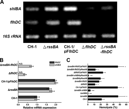
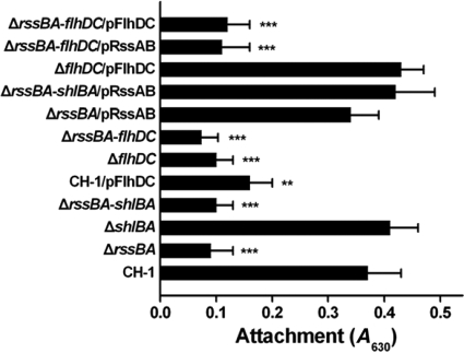
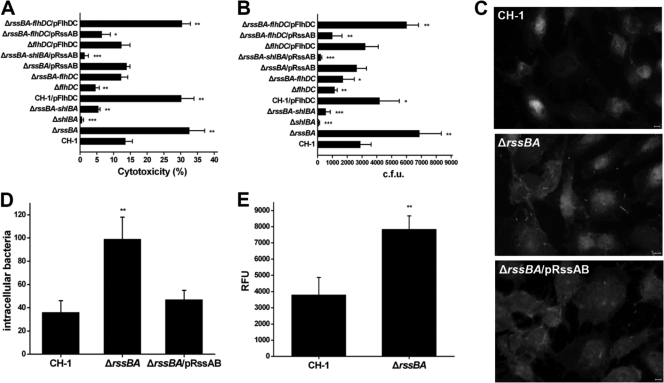
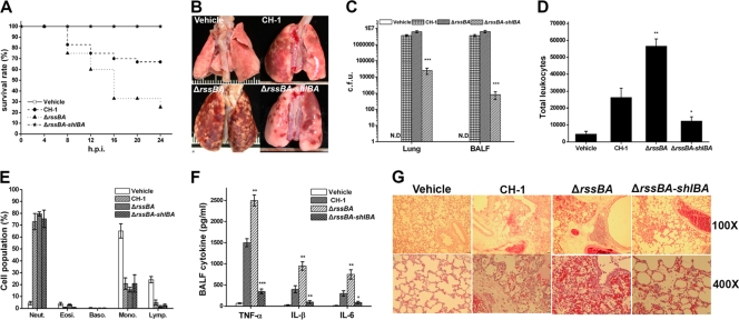
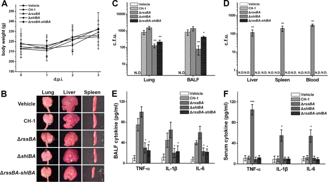
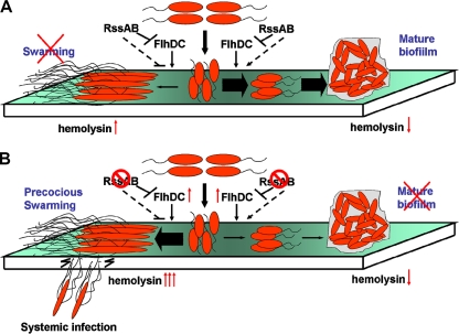
Serratia marcescens has long been recognized as an important opportunistic pathogen, but the underlying pathogenesis mechanism is not completely clear. Here, we report a key pathogenesis pathway in S. marcescens comprising the RssAB two-component system and its downstream elements, FlhDC and the dominant virulence factor hemolysin ShlBA. Expression of shlBA is under the positive control of FlhDC, which is repressed by RssAB signaling. At 37°C, functional RssAB inhibits swarming, represses hemolysin production, and promotes S. marcescens biofilm formation. In comparison, when rssBA is deleted, S. marcescens displays aberrant multicellularity favoring motile swarming with unbridled hemolysin production. Cellular and animal infection models further demonstrate that loss of rssBA transforms this opportunistic pathogen into hypervirulent phenotypes, leading to extensive inflammatory responses coupled with destructive and systemic infection. Hemolysin production is essential in this context. Collectively, a major virulence regulatory pathway is identified in S. marcescens.


Figures

FIG. 1.
FIG. 1.
RssAB represses hemolysin synthesis and hemolytic activity through downregulation of flhDC expression. (A) The mRNA expression levels of shlBA and flhDC in different S. marcescens strains grown to mid-log phase in LB broth at 37°C were determined by RT-PCR as shown on the agarose gel. (B) The transcript amount was semiquantified by Image J and is shown as relative mRNA expression, which is normalized to the 16S rRNA expression level and compared to that of S. marcescens CH-1. N.D., not detectable. (C) The percentage of relative hemolytic activity of each S. marcescens strain grown in LB broth at 37°C was determined by hemolysis assay. All results are shown as the average of three independent experiments, with the standard deviations indicated by error bars. *, P < 0.05, and ***, P < 0.001 in comparison to CH-1 (Student's t test).
FIG. 2.
FIG. 2.
RssAB and FlhDC are required for efficient biofilm attachment. Shown is the quantification of bacterial attachment efficacy by microtiter plate assay. The results were expressed as the A630. Expression of flhDC in S. marcescens CH-1/pFlhDC was induced by supplementation with 0.1% arabinose. All results are shown as the average of three independent experiments, with the standard deviation indicated by error bars. *, P < 0.05; **, P < 0.01; and ***, P < 0.001 in comparison to CH-1 (Student's t test).
FIG. 3.
FIG. 3.
Deletion of rssBA increases S. marcescens cellular cytotoxicity, invasive activity, and shlBA promoter activity. (A) Each S. marcescens strain was cultivated in LB broth to mid-log phase at 37°C before infection of the human bronchial epithelial cell line BEAS-2B at an MOI of 10 for 4 h. The percent cytotoxicity was determined by release of lactate dehydrogenase. (B) Invasion of BEAS-2B cells by different S. marcescens strains was determined by an antibiotic protection assay. The bacterial growth conditions were the same as those used in the cytotoxicity assay. The results are expressed as the average number of CFU from three independent experiments. (C) Each bacterial strain harboring pBAD24EGFP::Sm was cultivated in LB broth containing 0.1% arabinose to mid-log phase and harvested. Bacteria at an MOI of 5 were used to infect BEAS-2B cells cultured on a coverslip at 37°C for 2 h. The cells were washed twice with PBS and stained with 5 μM FM 4-64 and 10 μM Hoechst. Under fluorescence microscopy, bacteria were localized and imaged. Scale bars, 5 μm. (D) The bacteria invading cells were counted in 200 BEAS-2B cells. (E) The shlBA promoter activities of S. marcescens CH-1 and S. marcescens ΔrssBA that harbored pRepoEGFP or pRepoEGFP-shlB were measured by fluorometer while they were infecting BEAS-2B cells. The results are shown as RFU. The data are shown as the mean from three independent experiments, with the standard deviations indicated by error bars. *, P < 0.05; **, P < 0.01; and ***, P < 0.001 in comparison to CH-1 (Student's t test).
FIG. 4.
FIG. 4.
ShlBA under RssAB control plays an important role in S. marcescens acute-pneumonia pathogenesis. (A) Rats (n = 12 in each group for independent experiments) were i.t. inoculated with 1 × 10⁸ CFU of S. marcescens CH-1, S. marcescens ΔrssBA, and S. marcescens ΔrssBA-shlBA and vehicle. The survival rate was monitored every 4 h for 24 h postinfection (h.p.i.). (B) The whole lung of a rat was dissected for a gross view and imaged 24 h p.i. (C) Bacterial burdens from the lung and BAL fluid (BALF) at 24 h p.i. were analyzed by plate counting. The results are expressed as the average number of CFU per gram of lung and CFU per ml of BALF. N.D., not detectable. (D) Total leukocyte counts in BALF from each group were determined by hemocytometer analysis. (E) The cell differential count was determined and expressed as an average ratio of total leukocyte counts. (F) The supernatant of BALF from each group 1 day postinfection was collected for measurement of TNF-α, IL-1β, and IL-6, determined as pg/ml, by ELISA. (G) Dissected lungs fixed in 10% formalin for 24 h and paraffin embedded were cut as 4-μm sections for H&E staining. The images were analyzed and captured with a Leica DM 2500 under ×100 and ×400 magnification. Scale bars, 40 μm and 10 μm for ×100 and ×400, respectively. All results are shown as the average of three independent experiments, with the standard deviations indicated by error bars. *, P < 0.05; **, P < 0.01; and ***, P < 0.001 in comparison to CH-1 (Student's t test).
FIG. 5.
FIG. 5.
The rssBA deletion leads to systemic infection of S. marcescens in a sublethal-pneumonia model of immunocompetent rats. Rats (n = 6 in each group for independent experiments) were i.t. infected with 1 × 10⁵ CFU of S. marcescens CH-1, S. marcescens ΔrssBA, S. marcescens ΔshlBA, and S. marcescens ΔrssBA-shlBA cells for 3 days postinfection (d.p.i.). Normal saline was used as a vehicle control. (A) Body weight was monitored daily. (B) On the third day p.i., the rats were sacrificed, and organs, including lungs, livers, and spleens, were removed for gross morphology observation accompanied by collection of BALF and sera. (C and D) The bacterial loads of organ homogenates, sera, and BALF were determined as the average number of CFU per gram of organ and per ml of serum or BALF. N.D., not detectable. (E and F) BALF and sera from each group 3 days p.i. were utilized to measure TNF-α, IL-1β, and IL-6 as determined by ELISA. All results are shown as the average of three independent experiments, with the standard deviations indicated by error bars. *, P < 0.05, and **, P < 0.01 in comparison to CH-1 (Student's t test).
FIG. 6.
FIG. 6.
Proposed mechanisms by which RssAB controls multicellularity and pathogenesis in S. marcescens. S. marcescens utilizes RssAB to coordinate multicellular behaviors, such as biofilm formation and swarming motility, with the concomitant modulation of virulence and hemolysin production, by regulating flhDC expression at 37°C. (A) Activation of RssAB signaling reduces flhDC expression and hemolysin production and favors biofilm formation over swarming. (B) In the absence of RssAB, flhDC expression is increased. S. marcescens becomes hypervirulent, with increased hemolysin production accompanied by precocious swarming and defective biofilm formation. This gives S. marcescens the capability to cause disseminated infection. The dashed arrows represent unidentified determinants probably involved in the proposed model.

References

    1. Allison, C., N. Coleman, P. L. Jones, and C. Hughes. 1992. Ability of Proteus mirabilis to invade human urothelial cells is coupled to motility and swarming differentiation. Infect. Immun. 60:4740-4746. - PMC - PubMed
    1. Bubeck Wardenburg, J., and O. Schneewind. 2008. Vaccine protection against Staphylococcus aureus pneumonia. J. Exp. Med. 205:287-294. - PMC - PubMed
    1. Buffet-Bataillon, S., V. Rabier, P. Betremieux, A. Beuchee, M. Bauer, P. Pladys, E. Le Gall, M. Cormier, and A. Jolivet-Gougeon. 2009. Outbreak of Serratia marcescens in a neonatal intensive care unit: contaminated unmedicated liquid soap and risk factors. J. Hosp. Infect. 72:17-22. - PubMed
    1. Caiazza, N. C., J. H. Merritt, K. M. Brothers, and G. A. O'Toole. 2007. Inverse regulation of biofilm formation and swarming motility by Pseudomonas aeruginosa PA14. J. Bacteriol. 189:3603-3612. - PMC - PubMed
    1. Chang, A. C., and S. N. Cohen. 1978. Construction and characterization of amplifiable multicopy DNA cloning vehicles derived from the P15A cryptic miniplasmid. J. Bacteriol. 134:1141-1156. - PMC - PubMed

Publication types

MeSH terms

Substances

LinkOut - more resources