. 2010 Nov;78(11):4936-43.
doi: 10.1128/IAI.01118-09. Epub 2010 Aug 16.

Gelatinase contributes to the pathogenesis of endocarditis caused by Enterococcus faecalis

Affiliations

Gelatinase contributes to the pathogenesis of endocarditis caused by Enterococcus faecalis

Lance R Thurlow et al. Infect Immun. 2010 Nov.

Abstract

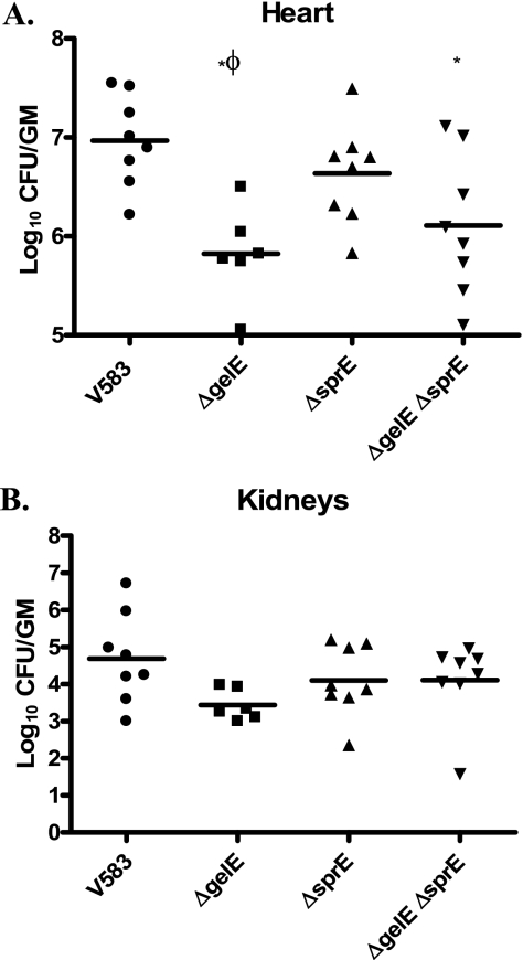
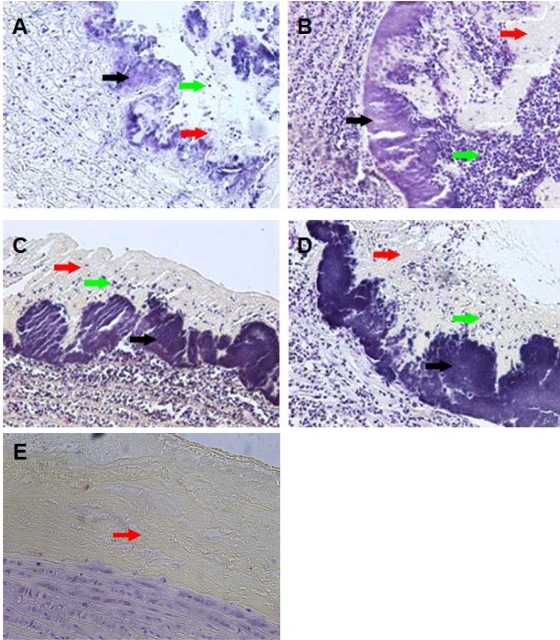
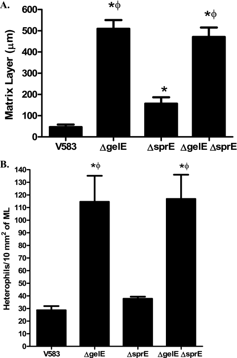
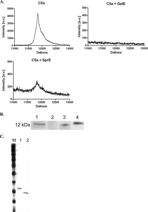
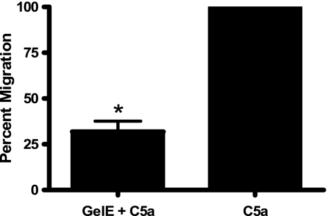
The Gram-positive pathogen Enterococcus faecalis is a leading agent of nosocomial infections, including urinary tract infections, surgical site infections, and bacteremia. Among the infections caused by E. faecalis, endocarditis remains a serious clinical manifestation and unique in that it is commonly acquired in a community setting. Infective endocarditis is a complex disease, with many host and microbial components contributing to the formation of bacterial biofilm-like vegetations on the aortic valve and adjacent areas within the heart. In the current study, we compared the pathogenic potential of the vancomycin-resistant E. faecalis V583 and three isogenic protease mutants (ΔgelE, ΔsprE, and ΔgelE ΔsprE mutants) in a rabbit model of enterococcal endocarditis. The bacterial burdens displayed by GelE(-) mutants (ΔgelE and ΔgelE ΔsprE mutants) in the heart were significantly lower than those of V583 or the SprE(-) mutant. Vegetations on the aortic valve infected with GelE(-) mutants (ΔgelE and ΔgelE ΔsprE mutants) also showed a significant increase in deposition of fibrinous matrix layer and increased chemotaxis of inflammatory cells. In support of a role for proteolytic modulation of the immune response to E. faecalis, we also demonstrate that GelE can cleave the anaphylatoxin complement C5a and that this proteolysis leads to decreased neutrophil migration in vitro. In vivo, a decreased heterophil (neutrophil-like cell) migration was observed at tissue sites infected with GelE-producing strains but not at those infected with SprE-producing strains. Taken together, these observations suggest that of the two enterococcal proteases, gelatinase is the principal mediator of pathogenesis in endocarditis.


Figures

FIG. 1.
FIG. 1.
Enterococcal burdens in the rabbit heart and kidneys. Vital organs of rabbits infected with E. faecalis (parental and isogenic protease mutants) were harvested following catheter-induced enterococcal endocarditis as described in Materials and Methods. (A) Mean bacterial burdens for V583 (parental), VT01 (ΔgelE), VT02 (ΔsprE), and VT03 (ΔgelE ΔsprE) are represented as log10 CFU/g of homogenized heart tissue. (B) Mean bacterial burdens in the pooled kidneys from each rabbit (n = 6 to 8). *, significant P values of less than 0.05 relative to V583; Φ, significant P values of less than 0.05 relative to the ΔsprE mutant (VT02).
FIG. 2.
FIG. 2.
Histology of aortic vegetations. Panels A, B, C, D, and E are representative images of Gram-stained cross-sections (5 μm) of vegetations formed on the ascending aorta of rabbits infected with V583, VT01 (ΔgelE), VT02 (ΔsprE), and VT03 (ΔgelE ΔsprE) and an uninfected control, respectively (magnification, ×200). Black arrows point to E. faecalis biomass on the surface of the endothelium. Red arrows point to deposited matrix layer composed mostly of platelets and fibrin. Green arrows point to influx of heterophils and other immune cell infiltrates.
FIG. 3.
FIG. 3.
(A) Matrix layer (ML) of animals infected with wild-type and extracellular protease mutants. Differences in the ML thickness were determined from histological images of vegetations (magnification, ×400). The lengths between the E. faecalis biomass layer and the upper edges of ML from eight random regions of vegetations from each strain were measured and reported as mean thickness (μm, mean ± standard error of the mean [SEM]). (B) Quantification of heterophil chemotaxis in the hearts of rabbits infected with E. faecalis. Differences in the numbers of heterophils that have migrated to the bacterial vegetations were determined from histological images (magnification, ×400) and were normalized to the area of ML surrounding them. Heterophils were counted using ImageJ software from four random images of vegetations from each strain and reported as the total number of heterophils trapped per 10 mm2 of ML (mean ± SEM). *, significant P values of less than 0.05 relative to V583; Φ, significant P values of less than 0.05 relative to the ΔsprE mutant (VT02).
FIG. 4.
FIG. 4.
GelE degrades C5a. (A) MALDI-TOF spectra of C5a (∼12 kDa) alone, C5a incubated with GelE, and C5a incubated with SprE. Incubation of C5a with GelE results in complete hydrolysis of C5a in 20 min, whereas incubation of C5a with SprE results in ∼ 90% hydrolysis under similar conditions. (B) Silver-stained Tris-Tricine gel showing the molecular mass marker of ∼12 kDa (lane 1), C5a incubated with GelE (lane 2), C5a incubated with SprE (lane 3), and C5a alone (lane 4). (C) Silver-stained gel of the purified proteases: M designates the molecular weight ladder. Lane 1, GelE; lane 2, SprE. The purified proteases were subjected to MALDI-TOF, and the molecular masses were determined to be 32,866.3 Da for GelE and 25,717.13 Da for SprE (data not shown).
FIG. 5.
FIG. 5.
Transwell migration assays. Incubation of C5a with GelE inhibits dHL-60 migration through Transwell membranes. Neutrophil-like dHL-60 cells were labeled with fluorogenic CFDA-SE and allowed to migrate through a 3.0-μm membrane in response to C5a or C5a previously incubated with GelE. Incubation of C5a with GelE significantly (*, P < 0.05) reduces dHL-60 chemotaxis compared to that of C5a alone.

References

    1. Cheng, Q., D. Stafslien, S. S. Purushothaman, and P. Cleary. 2002. The group B streptococcal C5a peptidase is both a specific protease and an invasin. Infect. Immun. 70:2408-2413. - PMC - PubMed
    1. Cleary, P. P., U. Prahbu, J. B. Dale, D. E. Wexler, and J. Handley. 1992. Streptococcal C5a peptidase is a highly specific endopeptidase. Infect. Immun. 60:5219-5223. - PMC - PubMed
    1. Daffern, P. J., P. H. Pfeifer, J. A. Ember, and T. E. Hugli. 1995. C3a is a chemotaxin for human eosinophils but not for neutrophils. I. C3a stimulation of neutrophils is secondary to eosinophil activation. J. Exp. Med. 181:2119-2127. - PMC - PubMed
    1. DiScipio, R. G., P. J. Daffern, M. A. Jagels, D. H. Broide, and P. Sriramarao. 1999. A comparison of C3a and C5a-mediated stable adhesion of rolling eosinophils in postcapillary venules and transendothelial migration in vitro and in vivo. J. Immunol. 162:1127-1136. - PubMed
    1. Engelbert, M., E. Mylonakis, F. M. Ausubel, S. B. Calderwood, and M. S. Gilmore. 2004. Contribution of gelatinase, serine protease, and fsr to the pathogenesis of Enterococcus faecalis endophthalmitis. Infect. Immun. 72:3628-3633. - PMC - PubMed

Publication types

MeSH terms

LinkOut - more resources