. 2010 Nov;78(11):4754-62.
doi: 10.1128/IAI.00789-10. Epub 2010 Aug 16.

The Inflammatory response induced by aspartic proteases of Candida albicans is independent of proteolytic activity

Affiliations

The Inflammatory response induced by aspartic proteases of Candida albicans is independent of proteolytic activity

Donatella Pietrella et al. Infect Immun. 2010 Nov.

Abstract

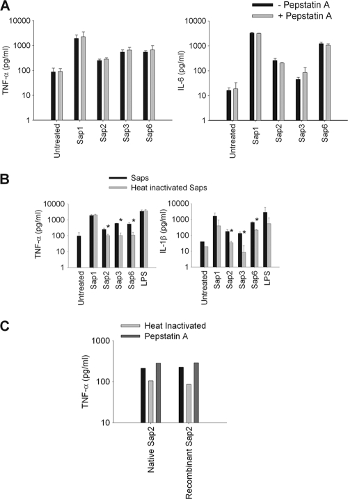
The secretion of aspartic proteases (Saps) has long been recognized as a virulence-associated trait of the pathogenic yeast Candida albicans. In this study, we report that different recombinant Saps, including Sap1, Sap2, Sap3, and Sap6, have differing abilities to induce secretion of proinflammatory cytokines by human monocytes. In particular Sap1, Sap2, and Sap6 significantly induced interleukin-1β (IL-1β), tumor necrosis factor alpha (TNF-α), and IL-6 production. Sap3 was able to stimulate the secretion of IL-1β and TNF-α. All Saps tested were able to induce Ca(2+) influx in monocytes. Treatment of these Saps with pepstatin A did not have any effect on cytokine secretion, indicating that their stimulatory potential was independent from their proteolytic activity. The capacity of Saps to induce inflammatory cytokine production was also independent from protease-activated receptor (PAR) activation and from the optimal pH for individual Sap activity. The interaction of Saps with monocytes induced Akt activation and phosphorylation of IκBα, which mediates translocation of NF-κB into the nucleus. Overall, these results suggest that individual Sap proteins can induce an inflammatory response and that this phenomenon is independent from the pH of a specific host niche and from Sap enzymatic activity. The inflammatory response is partially dependent on Sap denaturation and is triggered by the Akt/NF-κB activation pathway. Our data suggest a novel, activity-independent aspect of Saps during interactions of C. albicans with the host.


Figures

FIG. 1.
FIG. 1.
TNF-α, IL-6, and IL-1β production from monocytes after stimulation with Candida albicans rSaps. (A) Monocytes were treated with rSap1, rSap2, rSap3, and rSap6 (20 μg/ml), with lipase of M. furfur (20 μg/ml), with pepsin from porcine gastric mucosa, or with LPS (1 μg/ml) or were left untreated and then were incubated for 18 h at 37°C. After incubation, supernatants were recovered and tested for the presence of TNF-α, IL-6, and IL-1β. (B) Monocytes were treated with different concentrations of rSaps or pepsin, ranging from 1 to 50 μg/ml, or were left untreated and then were incubated for 18 h at 37°C. (C) Monocytes were treated with different rSaps (20 μg/ml) and LPS (1 μg/ml) in the presence or absence of 10 μg/ml of polymyxin B. (D) Monocytes were treated with native Sap2, rSap2, and culture supernatant. Data are expressed as means ± SD for three independent experiments. Statistical analysis was performed with an ANOVA test for multiple comparisons. *, P < 0.05; **, P < 0.01 (rSap-treated cells versus untreated cells).
FIG. 2.
FIG. 2.
Effects of heat inactivation of Saps, pepstatin A treatment, and anti-human PAR antibodies on cytokine production induced by Saps. Monocytes were treated with rSap1, rSap2, rSap3, and rSap6 (20 μg/ml) in combination with pepstatin A (15 μM) (A) or with heat-inactivated rSap1, rSap2, rSap3, rSap6, or LPS (1 μg/ml) (B). (C) Effect of pepstatin A (15 μM) or heat inactivation of native or recombinant Sap2 (20 μg/ml) on TNF-α production. After incubation, supernatants were recovered and tested for the presence of TNF-α, IL-1β, or IL-6. Data are expressed as means ± SD for three independent experiments. Statistical analysis was performed with an ANOVA test. *, P < 0.05 (heat-inactivated rSap-treated cells versus rSap-treated cells).
FIG. 3.
FIG. 3.
Association of FITC-Saps with monocytes. Monocytes treated with different concentrations of FITC-labeled rSap1 or rSap2 or with FITC-conjugated CTLA-4 F(ab)2 for different times were analyzed by flow cytometry to assess rSap binding. FITC-rSap or FITC-CTLA-4 F(ab)2 binding by monocytes is expressed as the mean fluorescence intensity (MIF). Data are expressed as means ± SD for three independent experiments.
FIG. 4.
FIG. 4.
pAkt and pIκB expression in monocytes treated with Saps. Monocytes were left untreated or were stimulated with rSap1, rSap2, rSap3, or Sap6 (20 μg/ml) (A and B) or with pepsin (20 μg/ml) (C) for 4 h. After incubation, cells were fixed in 3% paraformaldehyde, permeabilized in 0.1% saponin, and incubated with rabbit anti-human phospho-Akt or phospho-IκBα and with TRITC-conjugated goat anti-rabbit IgG. Fluorescent cells were analyzed by cytofluorimetric analysis and are expressed as the mean fluorescence intensity (MIF). Data are representative of three independent experiments with similar results.
FIG. 5.
FIG. 5.
Effect of Akt signaling inhibition on cytokine production induced by rSaps. Monocytes were left untreated or were pretreated with 5 μM wortmannin for 40 min and then treated with rSap1, rSap2, rSap3, or rSap6 (20 μg/ml) or with LPS (1 μg/ml) for 18 h at 37°C. After incubation, supernatants were recovered and tested for the presence of TNF-α, IL-6, and IL-1β. Data are expressed as means ± SD for three independent experiments. Statistical analysis was performed with an ANOVA test for multiple comparisons. *, P < 0.05; **, P < 0.01 (rSap-plus-wortmannin-treated cells versus rSap-treated cells).

Similar articles

Cited by

References

    1. Albrecht, A., A. Felk, I. Pichova, J. R. Naglik, M. Schaller, P. de Groot, D. Maccallum, F. C. Odds, W. Schafer, F. Klis, M. Monod, and B. Hube. 2006. Glycosylphosphatidylinositol-anchored proteases of Candida albicans target proteins necessary for both cellular processes and host-pathogen interactions. J. Biol. Chem. 281:688-694. - PubMed
    1. Borelli, C., E. Ruge, J. H. Lee, M. Schaller, A. Vogelsang, M. Monod, H. C. Korting, R. Huber, and K. Maskos. 2008. X-ray structures of Sap1 and Sap5: structural comparison of the secreted aspartic proteinases from Candida albicans. Proteins 72:1308-1319. - PubMed
    1. Borg-von Zepelin, M., S. Beggah, K. Boggian, D. Sanglard, and M. Monod. 1998. The expression of the secreted aspartyl proteinases Sap4 to Sap6 from Candida albicans in murine macrophages. Mol. Microbiol. 28:543-554. - PubMed
    1. Brunke, S., and B. Hube. 2006. MfLIP1, a gene encoding an extracellular lipase of the lipid-dependent fungus Malassezia furfur. Microbiology 152:547-554. - PubMed
    1. Calderone, R. A., and W. A. Fonzi. 2001. Virulence factors of Candida albicans. Trends Microbiol. 9:327-335. - PubMed

Publication types

MeSH terms