. 2010 Nov;54(11):4714-22.
doi: 10.1128/AAC.00974-10. Epub 2010 Aug 16.

1-O-hexadecyloxypropyl cidofovir (CMX001) effectively inhibits polyomavirus BK replication in primary human renal tubular epithelial cells

Affiliations

1-O-hexadecyloxypropyl cidofovir (CMX001) effectively inhibits polyomavirus BK replication in primary human renal tubular epithelial cells

Christine Hanssen Rinaldo et al. Antimicrob Agents Chemother. 2010 Nov.

Abstract

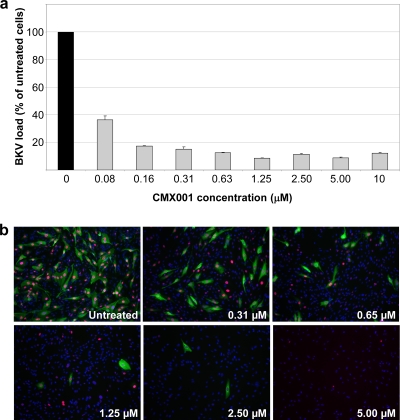
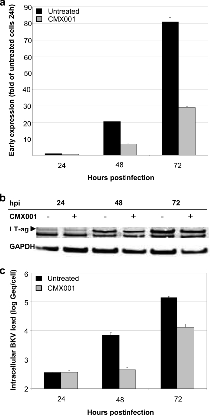
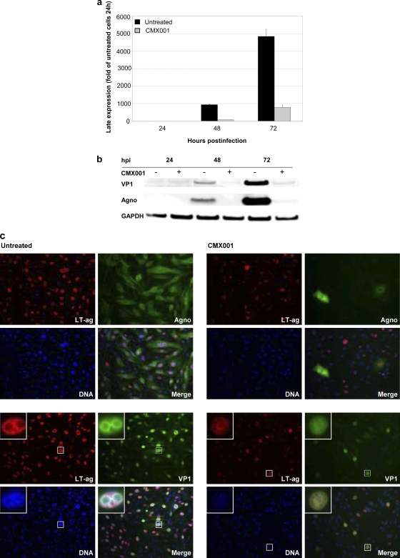
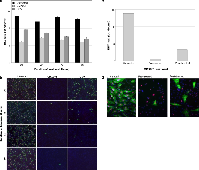
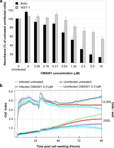
Antiviral drugs for treating polyomavirus BK (BKV) replication in polyomavirus-associated nephropathy or hemorrhagic cystitis are of considerable clinical interest. Unlike cidofovir, the lipid conjugate 1-O-hexadecyloxypropyl cidofovir (CMX001) is orally available and has not caused detectable nephrotoxicity in rodent models or human studies to date. Primary human renal proximal tubular epithelial cells were infected with BKV-Dunlop, and CMX001 was added 2 h postinfection (hpi). The intracellular and extracellular BKV DNA load was determined by quantitative PCR. Viral gene expression was examined by quantitative reverse transcription-PCR, Western blotting, and immunofluorescence microscopy. We also examined host cell viability, proliferation, metabolic activity, and DNA replication. The titration of CMX001 identified 0.31 μM as the 90% effective concentration (EC(90)) for reducing the extracellular BKV load at 72 hpi. BKV large T antigen mRNA and protein expression was unaffected at 24 hpi, but the intracellular BKV genome was reduced by 90% at 48 hpi. Late gene expression was reduced by 70 and 90% at 48 and 72 hpi, respectively. Comparisons of CMX001 and cidofovir EC(90)s from 24 to 96 hpi demonstrated that CMX001 had a more rapid and enduring effect on BKV DNA and infectious progeny at 96 hpi than cidofovir. CMX001 at 0.31 μM had little effect on overall cell metabolism but reduced bromodeoxyuridine incorporation and host cell proliferation by 20 to 30%, while BKV infection increased cell proliferation in both rapidly dividing and near-confluent cultures. We conclude that CMX001 inhibits BKV replication with a longer-lasting effect than cidofovir at 400× lower levels, with fewer side effects on relevant host cells in vitro.


Figures

FIG. 1.
FIG. 1.
Effect of increasing concentrations of CMX001 on BKV load and expression of BKV proteins. (a) RPTEC supernatants were harvested 72 hpi, i.e., 70 h after the start of treatment with indicated CMX001 concentrations, and BKV load was measured by qPCR. The DNA load in untreated cells (1.19 × 10⁹ genome equivalent [Geq]/ml) was set as 100%. Determinations were in triplicate. The mean values are shown, and the error bars represent standard deviations. (b) Indirect immunofluorescence of BKV-infected RPTECs either untreated or treated with the indicated CMX001 concentrations. The cells were methanol fixed 72 hpi and stained using as primary antibodies polyclonal rabbit anti-agno serum (green) for the visualization of the late agno and the SV40 LT-ag monoclonal Pab416 for the visualization of early LT-ag (red). Cell nuclei (blue) were stained with Drac 5.The pictures are taken with the 10× objective.
FIG. 2.
FIG. 2.
Influence of CMX001 at 0.31 μM on BKV-Dunlop early expression and DNA replication in RPTECs. (a) Early mRNA expression. RNA was extracted from CMX001-treated and untreated BKV-infected RPTECs at the indicated time points. LT-ag mRNA expression was measured by RT-qPCR and normalized to huHPRT transcripts. Results are presented as changes in the LT-ag mRNA level, with the level in the untreated sample at 24 hpi arbitrarily set to 1. Determinations were in triplicate. The mean values are shown, and the error bars represent standard deviations. (b) Early protein expression. Cell extracts from CMX001-treated (+) and untreated (−) BKV-infected RPTECs were harvested 24, 48, and 72 hpi, and Western blotting was performed with polyclonal rabbit anti-LT-ag serum and with a monoclonal antibody directed against the housekeeping protein glyceraldehyde-3-phosphate dehydrogenase (GAPDH). The anti-LT-ag serum also recognizes a cellular protein of unknown origin. (c) BKV DNA replication. CMX001-treated and untreated BKV-infected RPTECs were harvested at the indicated time points and DNA extracted. Intracellular BKV DNA load was measured by qPCR and normalized for cellular DNA using the aspartoacyclase (ACY) qPCR. Data are presented as log Geq/cell. Determinations were in triplicate. The mean values are shown, and the error bars represent standard deviations.
FIG. 3.
FIG. 3.
Influence of CMX001 at 0.31 μM on BKV-Dunlop late expression in RPTECs. (a) Late mRNA expression. RNA was extracted from CMX001-treated and untreated BKV-infected RPTECS at the indicated time points. VP1 mRNA expression was measured by RT-qPCR and normalized to huHPRT transcripts. Results are presented as changes in the VP1 mRNA level, with the level in the untreated sample at 24 hpi arbitrarily set to 1. Determinations were in triplicate. The mean values are shown, and the error bars represent standard deviations. (b) Late protein expression. Cell extracts from CMX001-treated (+) and untreated (−) BKV-infected RPTECs were harvested 24, 48, and 72 hpi, and Western blotting was performed with polyclonal rabbit anti-agno and anti-VP1 serum and with the monoclonal antibody anti-GAPDH. (c) Early and late protein expression. Indirect immunofluorescence of BKV-infected RPTECs either untreated or treated with CMX001. The cells were methanol fixed 72 hpi and stained using the primary antibodies polyclonal rabbit anti-agno serum or anti-VP1 serum (both green) for the visualization of the late agno and VP1 protein, respectively, in combination with the SV40 LT-ag monoclonal Pab416 for the visualization of early LT-ag (red). Cell nuclei (blue) were stained with Drac 5. The pictures are taken with the 20× objective. Inserts show selected BKV-infected cells.
FIG. 4.
FIG. 4.
Kinetics of CMX001 at 0.31 μM treatment of BKV-infected RPTECs. (a) Extracellular BKV load. Cells were infected for 2 h, and CMX001 was added for 22, 46, 70, or 94 h, respectively. At the indicated times, supernatant was removed, cells were washed, and new medium added. At 96 hpi all supernatants were harvested and qPCR was performed. Data are presented as BKV load in log Geq/ml. Determinations were in triplicate. The mean values are shown, and the error bars represent standard deviations. (b) Infection and indirect immunofluorescence. The supernatant collected 96 hpi from the cells described above where diluted 1:10 and seeded on new RPTEC cells. At 72 hpi cells were methanol fixed, and immunofluorescence staining with polyclonal rabbit anti-agno serum (green) and the SV40 LT-ag monoclonal Pab416 was performed (red). Cell nuclei (blue) were stained with Drac 5. The pictures were taken with the 10× objective. (c) Extracellular BKV load. CMX001 was added 24 h before infection was indicated. Before infection, all cells were washed once with complete medium. BKV was added for 2 h and then removed, and cells were washed once with medium. Where indicated, previously untreated cells now were treated with CMX001 for 24 h. After 24 h, all supernatants were removed and cells were washed once more before complete medium was given for 48 h. Supernatants were harvested 72 hpi, and BKV loads were measured by qPCR. Determinations were in triplicate. The mean values are shown, and the error bars represent standard deviations. (d) Indirect immunofluorescence of BKV-infected RPTECs treated as outlined above. The cells were methanol fixed and stained using the primary antibodies polyclonal rabbit anti-agno serum (green) for the visualization of the viral late gene protein agno and the SV40 LT-ag monoclonal Pab416 for the visualization of viral early protein LT-ag (red). Cell nuclei (blue) were stained with Drac 5. The pictures were taken with a 20× objective.
FIG. 5.
FIG. 5.
Influence of CMX001 on DNA replication, metabolic activity, cell adhesion, and the proliferation of uninfected and BKV-infected RPTECs. (a) Cellular DNA replication was examined with a cell proliferation enzyme-linked immunosorbent assay (ELISA) monitoring BrdU incorporation, and metabolic activity was examined with cell proliferation reagent WST-1 to measure WST-1 cleavage. Medium with indicated CMX001 concentrations was added 2 hpi, and absorbance was measured 72 hpi. Absorbance for untreated uninfected cells was set as 100%. Determinations were based on measurements of quintuplicates (5 wells). The mean values are shown, and the error bars represent standard deviations. (b) For a dynamic monitoring of cell adhesion and the proliferation of RPTECs, the XCELLigence system was used. RPTECs at a density of 2,000 and 12,000 cells/well were seeded on E plates. Twenty-seven hours after seeding, 150 μl of the medium in each well (a total of 200 μl) was replaced with fresh medium with or without purified BKV-Dunlop (MOI, 5) and with or without CMX001 (total concentration of 0.31 μM), and the cells were left until 96 h after cell seeding.

Similar articles

Cited by

References

    1. Bernhoff, E., T. J. Gutteberg, K. Sandvik, H. H. Hirsch, and C. H. Rinaldo. 2008. Cidofovir inhibits polyomavirus BK replication in human renal tubular cells downstream of viral early gene expression. Am. J. Transplant. 8:1413-1422. - PubMed
    1. Bernhoff, E., G. D. Tylden, L. J. Kjerpeseth, T. J. Gutteberg, H. H. Hirsch, and C. H. Rinaldo. 2010. Leflunomide inhibition of BK virus replication in renal tubular epithelial cells. J. Virol. 84:2150-2156. - PMC - PubMed
    1. Binet, I., V. Nickeleit, H. H. Hirsch, O. Prince, P. Dalquen, F. Gudat, M. J. Mihatsch, and G. Thiel. 1999. Polyomavirus disease under new immunosuppressive drugs: a cause of renal graft dysfunction and graft loss. Transplantation 67:918-922. - PubMed
    1. Binggeli, S., A. Egli, S. Schaub, I. Binet, M. Mayr, J. Steiger, and H. H. Hirsch. 2007. Polyomavirus BK-specific cellular immune response to VP1 and large T-antigen in kidney transplant recipients. Am. J. Transplant. 7:1131-1139. - PubMed
    1. Cole, C. N. 1996. Polyomavirinae: the viruses and their replication, p. 917-945. In D. M. K. Bernard, N. Fields, and Peter M. Howley (ed.), Fundamental virology, 3rd ed. Lippincott-Raven Publishers, Philadelphia, PA.

Publication types

MeSH terms

LinkOut - more resources