. 2010 Aug 31;107(35):15595-600.
doi: 10.1073/pnas.1001740107. Epub 2010 Aug 16.

Stabilization of neurotoxic Alzheimer amyloid-beta oligomers by protein engineering

Affiliations

Stabilization of neurotoxic Alzheimer amyloid-beta oligomers by protein engineering

Anders Sandberg et al. Proc Natl Acad Sci U S A. .

Abstract

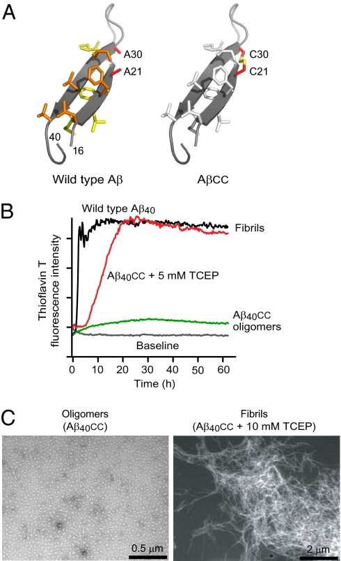
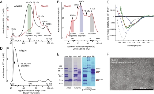
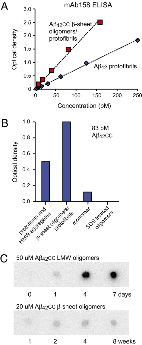
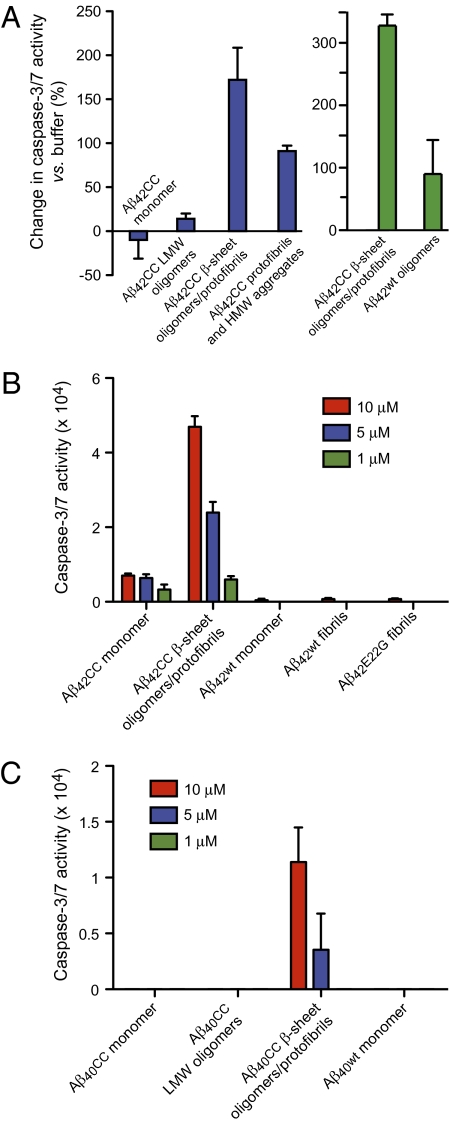
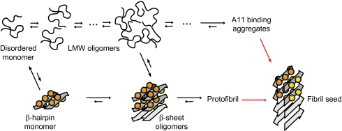
Soluble oligomeric aggregates of the amyloid-beta peptide (Abeta) have been implicated in the pathogenesis of Alzheimer's disease (AD). Although the conformation adopted by Abeta within these aggregates is not known, a beta-hairpin conformation is known to be accessible to monomeric Abeta. Here we show that this beta-hairpin is a building block of toxic Abeta oligomers by engineering a double-cysteine mutant (called Abetacc) in which the beta-hairpin is stabilized by an intramolecular disulfide bond. Abeta(40)cc and Abeta(42)cc both spontaneously form stable oligomeric species with distinct molecular weights and secondary-structure content, but both are unable to convert into amyloid fibrils. Biochemical and biophysical experiments and assays with conformation-specific antibodies used to detect Abeta aggregates in vivo indicate that the wild-type oligomer structure is preserved and stabilized in Abetacc oligomers. Stable oligomers are expected to become highly toxic and, accordingly, we find that beta-sheet-containing Abeta(42)cc oligomers or protofibrillar species formed by these oligomers are 50 times more potent inducers of neuronal apoptosis than amyloid fibrils or samples of monomeric wild-type Abeta(42), in which toxic aggregates are only transiently formed. The possibility of obtaining completely stable and physiologically relevant neurotoxic Abeta oligomer preparations will facilitate studies of their structure and role in the pathogenesis of AD. For example, here we show how kinetic partitioning into different aggregation pathways can explain why Abeta(42) is more toxic than the shorter Abeta(40), and why certain inherited mutations are linked to protofibril formation and early-onset AD.


Conflict of interest statement

Conflict of interest statement: A.S. and T.H. are shareholders of MIVAC Development AB, Gothenburg, Sweden.

Figures

Fig. 1.
Fig. 1.
Protein engineering. (A) (Left) The β-hairpin conformation of Aβ40 observed in complex with an Affibody binding protein (17). Nonpolar side chains at the two hydrophobic faces are shown as sticks and colored yellow and orange, respectively. The Ala21 and Ala30 methyls are located in close proximity on opposite β-strands. (Right) Model of the AβA21C/A30C double mutant (Aβcc) in which the β-hairpin conformation is locked by a disulfide bond. (B) ThT fluorescence assays of Aβ40cc aggregation in the absence or presence of TCEP reducing agent compared with wild-type Aβ40 aggregation. (C) TEM micrographs of β-sheet oligomers of Aβ40cc (Left) and of fibrils formed in presence of TCEP (Right).
Fig. 2.
Fig. 2.
Biophysical and biochemical characterization of Aβcc oligomers and protofibrils. (A) Formation and separation of Aβ42cc (black) and Aβ40cc (red) oligomers during SEC on a Superdex 200 PG 16/600 column. Monomer peptide samples were loaded in denaturing buffer and eluted with native phosphate buffer at pH 7.2. Apparent molecular weights and classification of eluted oligomers have been indicated. HMW aggregates elute with the void volume. Sample amounts: 2.7 mg Aβ40cc and 1.7 mg Aβ42cc; see also Fig. S2. (B) SEC as in A showing separation of Aβ40cc oligomers on a Superdex 75 PG 16/60 column on which smaller aggregates become better separated. Sample amount: ∼ 3 mg. (C) CD (mean residue ellipticity) of SEC fractions pooled as indicated by shaded areas in A and B. Dashed lines: 8-kDa monomer (gray, 12 μM), 30-kDa LMW oligomers (blue, 16 μM), and 96-kDa β-sheet oligomers (red, 8 μM) of Aβ40cc. Green: ∼100-kDa β-sheet oligomers of Aβ42cc (13 μM). Solid blue line: the 30-kDa LMW oligomer fraction of Aβ40cc after concentration and heat treatment showing formation of β-sheet oligomers. (D) SEC of concentrated Aβ42cc β-sheet oligomers (1 mL, 145 μM), which form protofibrils with an average apparent molecular weight of ∼ 650 kDa. The dotted line shows that these are smaller than HMW aggregates in A. (E) SDS/PAGE of wild-type Aβ42 (Left), purified but unfractionated Aβ42cc (Center), and Aβ42cc β-sheet oligomers formed during SEC as in A (Right). The right lanes in all panels contain Aβ samples, and other lanes contain high and low molecular weight standards (HMW and LMW). Weights corresponding to monomer and SDS-resistant dimers and trimers have been indicated. The loading buffer contained 2.5 mM TCEP (heat-stable reducing agent) to completely break all disulfide bonds. (F) TEM micrograph of a concentrated sample of Aβ42cc β-sheet oligomers (190-μM monomer concentration) showing assembly of β-sheet oligomers into protofibrils.
Fig. 3.
Fig. 3.
Recognition of Aβcc oligomers by antibodies that are conformation specific for wild-type Aβ aggregates. (A) mAb158 antibody sandwich ELISA detection (22) of Aβ42cc β-sheet oligomers/protofibrils compared with detection of wild-type Aβ42 protofibrils. (B) mAb158 ELISA analysis of different Aβ42cc species at 83 pM total peptide concentrations. (C) Aging of Aβ42cc LMW and β-sheet oligomers at 37 °C followed by A11 antibody dot blot analysis showing that A11 binding aggregates form from LMW oligomers but not from β-sheet oligomers. Concentrations were equalized before blotting.
Fig. 4.
Fig. 4.
Neurotoxicity of Aβcc to SH-SY5Y human neuroblastoma cells. Aβcc samples, except β-sheet oligomers/protofibrils in C, were prepared and isolated by SEC, as in Fig 2A, concentrated to 75 to 250 μM in phosphate buffer at pH 7.2, and added to cell cultures at 1-, 5-, or 10-μM concentrations. Caspase-3/7 activity reporting on apoptosis was measured after 24 h of treatment. (A) Aβ42cc induced apoptosis following treatment with 10 μM of different species. Blue: comparison of different Aβ42cc species. Green: Aβ42cc oligomers compared with wild-type Aβ42 oligomers in another experiment. (B) Dose-dependence of apoptosis induced by Aβ42cc species compared with that of wild-type Aβ42 monomer and fibrils and Aβ42E22G. (C) Apoptosis induced by different Aβ40cc species and wild-type Aβ40. The sample marked “Aβ40cc LMW oligomers” is a 75-kDa SEC oligomer fraction. The Aβ40cc β-sheet oligomers/protofibrils were formed by pooling and concentrating monomer and LMW oligomer SEC fractions and heating the concentrated sample as described in the text and in Fig. S4.
Fig. 5.
Fig. 5.
Aβ aggregation via two pathways. One pathway involves LMW oligomers without regular secondary structure and eventually large nonfibrillar aggregates binding the A11 antibody (coil pathway; Upper) and the other involves assembly into β-sheet oligomers, or coil oligomers that are converted into β-sheet oligomers, which are building blocks of mAb158 binding protofibrils (β-sheet pathway; Lower). Neurotoxic Aβ aggregates are formed along the β-sheet pathway. The scheme is consistent with the present studies of Aβcc and overall features can be reconciled with a large body of work on wild-type and naturally occurring Aβ mutants as discussed in the text. Red arrows reflect the interconversion of Aβ subunits from β-hairpin conformation in soluble aggregates to cross-β structure in fibril seeds and mature amyloid fibrils.

References

    1. Chiti F, Dobson CM. Protein misfolding, functional amyloid, and human disease. Annu Rev Biochem. 2006;75:333–366. - PubMed
    1. Roychaudhuri R, Yang M, Hoshi MM, Teplow DB. Amyloid beta-protein assembly and Alzheimer disease. J Biol Chem. 2009;284:4749–4753. - PMC - PubMed
    1. Shankar GM, et al. Amyloid-beta protein dimers isolated directly from Alzheimer’s brains impair synaptic plasticity and memory. Nat Med. 2008;14:837–842. - PMC - PubMed
    1. Lesné S, et al. A specific amyloid-β protein assembly in the brain impairs memory. Nature. 2006;440:352–357. - PubMed
    1. De Felice FG, et al. Abeta oligomers induce neuronal oxidative stress through an N-methyl-D-aspartate receptor-dependent mechanism that is blocked by the Alzheimer drug memantine. J Biol Chem. 2007;282:11590–11601. - PubMed

Publication types

MeSH terms

LinkOut - more resources