. 2010 Aug 31;107(35):15577-82.
doi: 10.1073/pnas.1009324107. Epub 2010 Aug 16.

Uterine FK506-binding protein 52 (FKBP52)-peroxiredoxin-6 (PRDX6) signaling protects pregnancy from overt oxidative stress

Affiliations

Uterine FK506-binding protein 52 (FKBP52)-peroxiredoxin-6 (PRDX6) signaling protects pregnancy from overt oxidative stress

Yasushi Hirota et al. Proc Natl Acad Sci U S A. .

Abstract

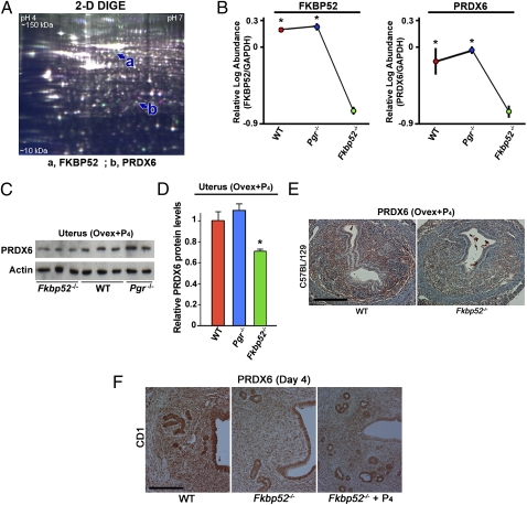
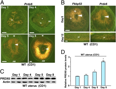
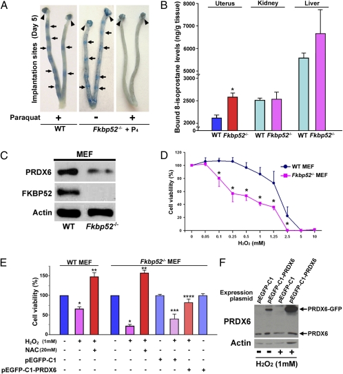
Immunophilin FK506-binding protein 52 (FKBP52) is a cochaperone that binds to the progesterone receptor (PR) to optimize progesterone (P(4))-PR signaling. We recently showed that Fkbp52-deficient (Fkbp52(-/-)) mice have reduced uterine PR responsiveness and implantation failure which is rescued by excess P(4) supplementation in a genetic background-dependent manner. This finding led us to hypothesize that FKBP52 has functions in addition to optimizing PR activity. Using proteomics analysis, we found that uterine levels of peroxiredoxin-6 (PRDX6), a unique antioxidant, are significantly lower in Fkbp52(-/-) mice than in WT and PR-null (Pgr(-/-)) mice. We also found that Fkbp52(-/-) mice with reduced uterine PRDX6 levels are susceptible to paraquat-induced oxidative stress (OS), leading to implantation failure even with P(4) supplementation. The same dose of paraquat did not interfere with implantation in WT mice. Moreover, treatment with antioxidants alpha-tocopherol and N-acetylcysteine (NAC) attenuated paraquat-induced implantation failure in P(4)-treated Fkbp52(-/-) mice. Functional analyses using mouse embryonic fibroblasts show that Fkbp52 deficiency associated with reduced PRDX6 levels promotes H(2)O(2)-induced cell death, which is reversed by the addition of NAC or by forced expression of PRDX6, suggesting that Fkbp52 deficiency diminishes the threshold against OS by reducing PRDX6 levels. These findings provide evidence that heightened uterine OS in Fkbp52(-/-) females with reduced PRDX6 levels induces implantation failure even in the presence of excess P(4). This study shows that FKBP52-PRDX6 signaling protects pregnancy from overt OS.


Conflict of interest statement

The authors declare no conflict of interest.

Figures

Fig. 1.
Fig. 1.
PRDX6 expression is reduced in uteri of Fkbp52−/− mice. (A) A representative DIGE gel comprising protein extracts from WT and Fkbp52−/− uteri shows proteins of isoelectric points between pH 4–7 and apparent molecular mass between 10–150 kDa. a, FKBP52; b, PRDX6. (B) Decreased protein levels of PRDX6 and FKBP52 in P4-treated uteri of Fkbp52−/− ovariectomized mice (n = 4) compared with WT (n = 3) and Pgr−/− (n = 3) ovariectomized mice on a C57BL6/129 background. Values are mean ± SEM. *P < 0.05 compared with Fkbp52−/− mice. (C and D) Decreased PRDX6 protein levels in P4-treated uteri of Fkbp52−/− ovariectomized mice compared with WT and Pgr−/− ovariectomized females as determined by Western blotting. Band intensities are normalized against actin and expressed as relative ratios compared with WT samples on the C57BL6/129 background. Values are mean ± SEM of two or three independent samples. *P < 0.05 compared with WT mice. (E) Differential expression patterns of PRDX6 protein in P4-treated ovariectomized mouse uteri of WT and Fkbp52−/− mice on the C57BL6/129 background. (Scale bar, 200 μm.) (F) Differential expression patterns of PRDX6 protein in day 4 uteri of WT and Fkbp52−/− mice on a CD1 background. (Scale bar, 200 μm.)
Fig. 2.
Fig. 2.
Uterine PRDX6 is expressed differentially during early pregnancy. (A) In situ hybridization showing spatiotemporal expression of Prdx6 in WT CD1 uteri on days 1, 4, 5, and 8 of pregnancy. Arrowheads indicate implanting embryos. AM, antimesometrial pole; ge, glandular epithelium; le, luminal epithelium; M, mesometrial pole; s, stroma. (Scale bar, 200 μm.) (B) Overlapping expression of Fkbp52 and Prdx6 in CD1 WT uteri on days 5 and 12 of pregnancy. Arrowheads indicate implanting embryos. le, luminal epithelium; pl, placenta; s, stroma. (Scale bars, 200 μm.) (C and D) Levels of uterine PRDX6 protein during early pregnancy as determined by Western blotting. Band intensities of PRDX6 were normalized against actin. Two independent samples from different mice were examined in each group. Data are expressed as mean ± SEM. *P < 0.05 compared with day 1 uteri.
Fig. 3.
Fig. 3.
Fkbp52−/− uteri are more prone to OS. (A) OS inducer paraquat blocks embryo implantation in P4-primed CD1 Fkbp52−/− females on day 5 of pregnancy, whereas paraquat-treated CD1 WT uteri have normal numbers of implantation sites as demarcated by blue bands. Fkbp52−/− females were treated with P4 (2 mg/d) on days 2–4 of pregnancy. Paraquat was injected on day 4. Arrows and arrowheads indicate implantation sites and ovaries, respectively. (B) Increased levels of bound 8-Isoprostane, a lipid peroxidation marker, in C57BL6/129 Fkbp52−/− uteri on day 4 of pseudopregnancy. Values are mean ± SEM of three independent samples. *P < 0.05 compared with WT mice. (C) Decreased levels of PRDX6 in Fkbp52−/− MEFs. Actin was used as a loading control. (D) Heightened sensitivity of Fkbp52−/− MEFs to H2O2-induced OS. Cells were treated with different concentrations of H2O2 for 24 h, and cell viability was evaluated by MTT assay. Values are mean ± SEM of six replicates from three independent experiments. *P < 0.05 compared with WT mice. (E) H2O2-induced cell death in Fkbp52−/− MEFs is protected by addition of NAC and forced expression of PRDX6. Cell viability was evaluated by MTT assay. Values are mean ± SEM of six replicates from three independent experiments. *P < 0.05, compared with vehicle-treated cells derived from mice of the same genotype. **P < 0.05, compared with H2O2-treated cells derived from mice of the same genotype. ***P < 0.05, compared with Fkbp52−/− cells transfected with pEGFP-C1 control plasmids; ****P < 0.05 compared with H2O2-treated Fkbp52−/− cells with transfection of pEGFP-C1 control plasmids. (F) PRDX6-GFP fused protein is induced effectively by transfection of pEGFP-C1-PRDX6 plasmids in Fkbp52−/− MEFs. pEGFP-C1 is a control plasmid.

Similar articles

Cited by

References

    1. Margalioth EJ, Ben-Chetrit A, Gal M, Eldar-Geva T. Investigation and treatment of repeated implantation failure following IVF-ET. Hum Reprod. 2006;21:3036–3043. - PubMed
    1. Dey SK, et al. Molecular cues to implantation. Endocr Rev. 2004;25:341–373. - PubMed
    1. Lydon JP, et al. Mice lacking progesterone receptor exhibit pleiotropic reproductive abnormalities. Genes Dev. 1995;9:2266–2278. - PubMed
    1. Smith DF. Tetratricopeptide repeat cochaperones in steroid receptor complexes. Cell Stress Chaperones. 2004;9:109–121. - PMC - PubMed
    1. Zhong L, et al. Heat-shock treatment-mediated increase in transduction by recombinant adeno-associated virus 2 vectors is independent of the cellular heat-shock protein 90. J Biol Chem. 2004;279:12714–12723. - PMC - PubMed

Publication types

MeSH terms

LinkOut - more resources