. 2010 Sep 15;185(6):3326-36.
doi: 10.4049/jimmunol.1000802. Epub 2010 Aug 16.

Ethylenecarbodiimide-treated splenocytes carrying male CD4 epitopes confer histocompatibility Y chromosome antigen transplant protection by inhibiting CD154 upregulation

Affiliations

Ethylenecarbodiimide-treated splenocytes carrying male CD4 epitopes confer histocompatibility Y chromosome antigen transplant protection by inhibiting CD154 upregulation

Aaron J Martin et al. J Immunol. .

Abstract

In humans and certain strains of laboratory mice, male tissue is recognized as nonself and destroyed by the female immune system via recognition of histocompatibility Y chromosome Ag (Hya). Male tissue destruction is thought to be accomplished by CTLs in a helper-dependent manner. We show that graft protection induced with the immunodominant Hya-encoded CD4 epitope (Dby) attached to female splenic leukocytes (Dby-SPs) with the chemical cross-linker ethylenecarbodiimide significantly, and often indefinitely, prolongs the survival of male skin graft transplants in an Ag-specific manner. In contrast, treatments with the Hya CD8 epitopes (Uty-/Smcy-SPs) failed to prolong graft survival. Dby-SP-tolerized CD4(+) T cells fail to proliferate, secrete IFN-gamma, or effectively prime a CD8 response in recipients of male grafts. Ag-coupled splenocyte treatment is associated with defective CD40-CD40L interactions as demonstrated by the observation that CD4 cells from treated animals exhibit a defect in CD40L upregulation following in vitro Ag challenge. Furthermore, treatment with an agonistic anti-CD40 Ab at the time of transplantation abrogates protection from graft rejection. Interestingly, anti-CD40 treatment completely restores the function of Dby-specific CD4 cells but not Uty- or Smcy-specific CD8 cells.


Figures
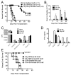
Figure 1
Figure 1. Hya-specific MHC class II, but not MHC class I, restricted Hya epitopes ECDI coupled to B6 female-derived splenocytes promote long-term, antigen-specific survival of male skin grafts on B6 female recipients
(A) Naïve C57BL/6 females were treated i.v. with syngeneic female splenic leukocytes ECDI-linked to the CD4 epitope (Dby) or the CD8 Hya epitopes (Uty and Smcy) or not tolerized on days -7 and 0 relative to engraftment with male tail skin grafts (and female control grafts – data not shown). Untreated control mice were included as a baseline for rejection time. Graft survival was monitored visually for 100 days. Male skin graft survival was significantly prolonged (*p<0.001) in female recipients treated with female Dby-SP (filled circles) compared to both untreated (open circles) and Uty/Smcy-SP treated (filled triangles) female recipients. (B) DTH responses of female B6 mice to ear challenge with 10μg soluble Dby were determined at 10, 20 and 60 days post transplantation in naïve, untreated and Dby-SP treated female B6 mice. Ear swelling responses in naïve C57BL/6 mice served as the baseline. Ear swelling responses in Dby-SP treated mice were significantly (*p<0.05) less than those in non-tolerized controls. In vitro recall responses of splenic T cells from untreated and Dby-SP tolerized mice were determined 10 days following transplantation upon stimulation with anti-CD3 (clone 2C11-positive control), OVA323-339 (negative control), and Dby peptides by [3H]-thymidine incorporation (C) and IFN-γ secretion (D). Proliferative and IFN-γ responses were significantly (*p <0.01) suppressed in Dby-SP tolerized animals upon challenge with either the CD4 (Dby) or CD8 (Uty and Smcy) Hya epitopes. Proliferative responses to Dby could be restored to control levels by the addition of 25 U/ml exogenous IL-2. (E) Antigen-specificity of the induction and effector stages of the regulatory effect was tested by treating female B6 mice i.v. with syngeneic female splenocytes ECDI-coupled with either OVA323-339 or the Dby peptide at days -7 and 0 relative to engraftment with both male B6 skin and female skin from a C57BL/10 donor. Neither Dby-SP (open circles) nor OVA323-339-SP (open triangles) treatment significantly prolonged B10 skin graft survival. However, Dby-SP (closed circles), but not OVA323-339-SP (closed triangles) significantly protected (*p<0.001) male B6 grafts from rejection. Five recipients were included in each group. All data shown represent at least 3 independent experiments.
Figure 2
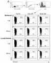
Figure 2. Dby-SP-induced nonresponsivenss of Hya-specific CD4+ T cells leads to failed priming Hya-specific CD8+ T cells specific for the Smcy and Uty epitopes
The activation frequency (CD44+) of Hya Uty- and Smcy-epitope-specific CD8+ T cells was determined using MHC Class-I tetramers. (A) Live, single CD8+ cells were identified. CD44 expression on Uty-specific CD8+ T cells in spleens (B-D) and draining LNs (H-J), and on Smcy-specific CD8+ T cells in spleens (E-G) and draining LNs (K-M) of naïve, non-tolerized (No Rx) and i.v. Dby-SP tolerized female B6 mice receiving B6 male tail skin grafts 14 d previously are shown. The total number of tetramer+ events is listed on each dot plot. Percentages of activated (CD44+) graft-specific CD8 cells, which appear in the top region of the tetramer+ box are also listed. Data are representative of three independent experiments.
Figure 3
Figure 3. Dby-SP-induced nonresponsivenss of Hya-specific CD4+ T cells results in diminished cytolytic activity of Hya-specific CD8+ T cells specific for the Smcy and Uty epitopes
B6 females were ungrafted (naïve), untreated (No Rx) or i.v. tolerized with Dby-SP prior to engraftment with male skin. 14 days later, in vivo Hya-specific cytolytic activity was determined. Peptide-loaded targets were administered (one specific target and one reference target) and were discernable by differential CFSE labeling. In panels A-D, whole male splenocytes (specific) vs. female splenocytes (reference) were used; in panels E-H, female splenocytes were pulsed with a combination of the Hya Uty and Smcy peptides (specific) or TMEV VP2 (reference); in panels I-J, female splenocytes were pulsed with the combination of the Hya Uty, Smcy, and Dby peptides (specific) or TMEV VP2 and OVA323-339 (reference). White bars on the graphs (Panels D, H, and L) represent calculated lysis in non-treated graft recipients, black bars represent lysis observed in Dby-SP-treated recipients, and grey bars represent lysis observed in unmanipulated naïve female controls. In vivo cytolytic responses in Dby-SP tolerized mice were significantly (*p<0.01) lower than those in non-tolerized controls. Data are representative of 3 independent experiments.
Figure 4
Figure 4. Ag-SP treatment inhibits CD154 upregulation on antigen-specific CD4+ T cells upon antigen recall
B6 OVA323-339-specific OTII TCR transgenic mice were untreated (naïve) (A&D) or injected i.v. with 108 OVA323-339-SP (B&E), or 108 MBP84-104-SP (C&F). Seven days later, splenocytes from these animals were harvested, stained and analyzed for surface expression of CD154 immediately upon explant and again at 6 hours following in vitro re-stimulation with 5μM OVA323-339. Maximal CD154 expression was observed at 6 h post-culture. FMO (G) = fluorescence minus one control. Data are representative of at least 3 independent experiments. 10,000 CD4+ events appear in each dot plot.
Figure 5
Figure 5. Dby-SP-induced protection of Hya skin grafts is reversed by CD40 cross-linking
(A) 5-7 untreated (No Rx) and Dby-SP tolerized female B6 mice received male tail skin grafts on day 0. 24 hours later, separate groups of treated mice were injected i.p. with 100μg of IgG2a isotype control antibody or with the agonistic anti-CD40 monoclonal antibody, FGK45.5. Graft survival was monitored by visual inspection for 100 days. The prolonged survival of male skin grafts observed in Dby-SP tolerized recipients treated with isotype control antibody was completely reversed by FGK45.5 treatment, *p<0.001. 10 days following transplantation and antibody treatment, recall responses of splenic T cells from the various treatment groups to in vitro stimulation with the Dby peptide were determined by [3H]-thymidine incorporation (B) and IFN-γ secretion (C). Suppressed proliferative and IFN-γ responses in the Dby-SP tolerized animals were significantly reversed by treatment with the FGK45.5, *p <0.01. Data is representative of at least 3 independent experiments.
Figure 6
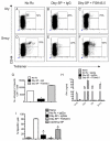
Figure 6. FGK45.5-mediated reversal of Dby-SP-induced protection of Hya skin grafts is not associated with restoration of Hya-specific CD8 T cell IFN-γ production or CTL activity
Untreated (No Rx) and Dby-SP tolerized female B6 mice received male tail skin grafts on day 0. 24 hours later, separate groups of Dby-SP treated mice were injected i.p. with 100 μg of IgG2a isotype control or with the agonistic anti-CD40 monoclonal antibody, FGK45.5. 10 days post-transplantation, spleens from these treated mice were analyzed for the frequency of activated (CD44+), Hya Uty- (A-C) and Smcy-epitope-specific CD8+ T cells (D-F) using MHC Class-I tetramers. (G) The total numbers of activated Hya-specific CD8+ T cells are plotted. Similar results were observed upon analysis of LN CD8+ T cells (not shown). The in vitro INF-γ recall response of splenocytes from the various treatment groups to recall stimulation with the Hya CD4 and CD8 epitopes was determined (H), as was the in vivo lytic activity of to targets pulsed with a combination of the Hya Uty and Smcy CD8 epitopes (target) or TMEV VP2 (reference) (I). Interestingly, the IFN-γ response was restored to stimulation with the CD4 Dby epitope, but neither IFN-γ production by nor the lytic function of CD8 T cells specific for the Uty or Smcy epitopes were restored by CD40 ligation. Data represent 2 independent experiments.
Figure 7
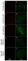
Figure 7. Reduced CD8 graft infiltrate does not correlate with graft survival
B6 female mice received female (panels A & B) and male (C-J) skin grafts following treatment with Uty/Smcy-SP (C & D), nothing (E & F), Dby-SP + rIgG2a (G & H), or Dby-SP + FGK45.5 (I & J). Ten days after engraftment, histologic sections were prepared from graft-containing tail areas and stained for CD4 (B, D, F, H, J) or CD8 (A, C, E, G, I). Grafts from mice displaying rejection (i.e. Uty/Smcy-SP-treated, Dby-SP + FGK45.5-treated, and untreated groups) contain variable amounts of CD8 infiltrate and considerable CD4 infiltrate. Grafts that are retained (female control grafts and male grafts on Dby-SP + rIgG2a-treated females) contain very few infiltrating T cells.
Figure 8
Figure 8. FGK45.5 treatment results in increased numbers of activated B cells and dendritic cells
C57BL/6 females were engrafted with skin from a CD45.1 congenic donor female. 24 hours following transplantation, recipient animals received 100μg of FGK45.5 or rat IgG2a via i.p. injection. 48 hours following antibody treatment (and 72h post-transplantation), spleens and lymph nodes were analyzed for the expression of the B7-family costimulatory molecule CD86 on various lineages of APCs. CD86 expression was upregulated on the cells of graft recipients treated with FGK45.5 (Panel D), but not those treated with isotype control (Panel C), nor on naïve control animals (B). Enumeration and phenotyping revealed that the majority of CD86 expressers are B cells (E) and dendritic cells (F). Three mice were included in each group.

References

    1. Miller SD, Turley DM, Podojil JR. Antigen-specific tolerance strategies for the prevention and treatment of autoimmune disease. Nat. Rev. Immunol. 2007;7:665–677. - PubMed
    1. Gonsette RE. Compared benefit of approved and experimental immunosuppressive therapeutic approaches in multiple sclerosis. Expert. Opin. Pharmacother. 2007;8:1103–1116. - PubMed
    1. Moreau T, Coles A, Wing M, Isaacs J, Hale G, Waldmann H, Compston A. Transient increase in symptoms associated with cytokine release in patients with multiple sclerosis. Brain. 1996;119:225–237. - PubMed
    1. Miller SD, Wetzig RP, Claman HN. The induction of cell-mediated immunity and tolerance with protein antigens coupled to syngeneic lymphoid cells. J. Exp. Med. 1979;149:758–773. - PMC - PubMed
    1. Vanderlugt CL, Eagar TN, Neville KL, Nikcevich KM, Bluestone JA, Miller SD. Pathologic role and temporal appearance of newly emerging autoepitopes in relapsing experimental autoimmune encephalomyelitis. J. Immunol. 2000;164:670–678. - PubMed

Publication types

MeSH terms