. 2010 Nov 11;116(19):3944-54.
doi: 10.1182/blood-2010-03-267419. Epub 2010 Aug 16.

Eosinophils in the zebrafish: prospective isolation, characterization, and eosinophilia induction by helminth determinants

Affiliations

Eosinophils in the zebrafish: prospective isolation, characterization, and eosinophilia induction by helminth determinants

Keir M Balla et al. Blood. .

Abstract

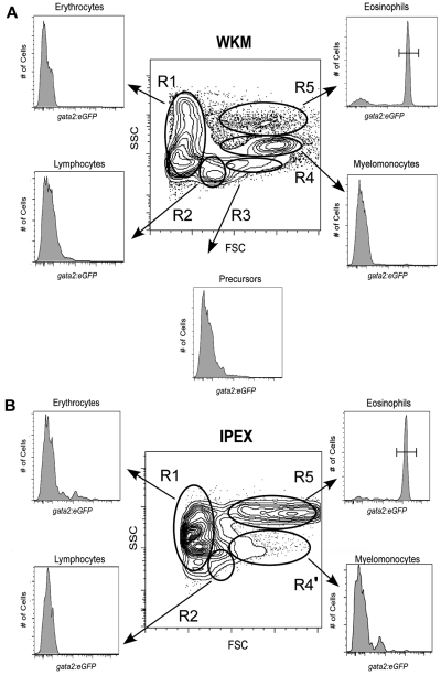
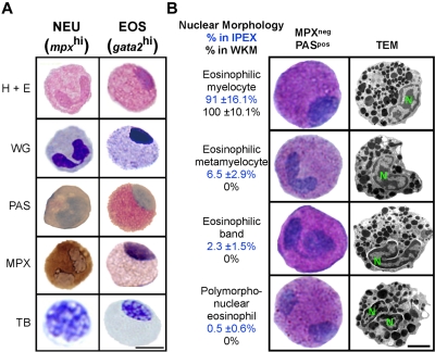
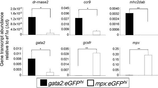
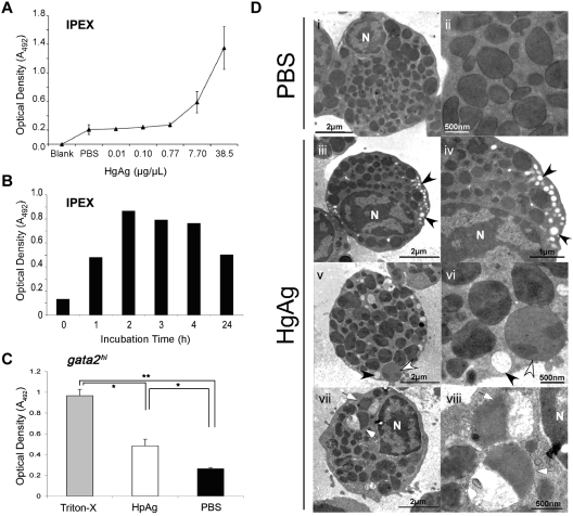
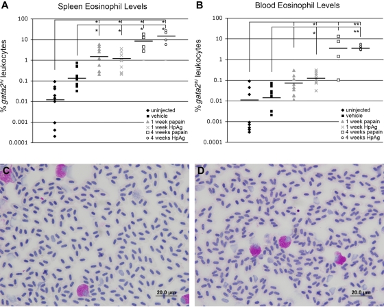
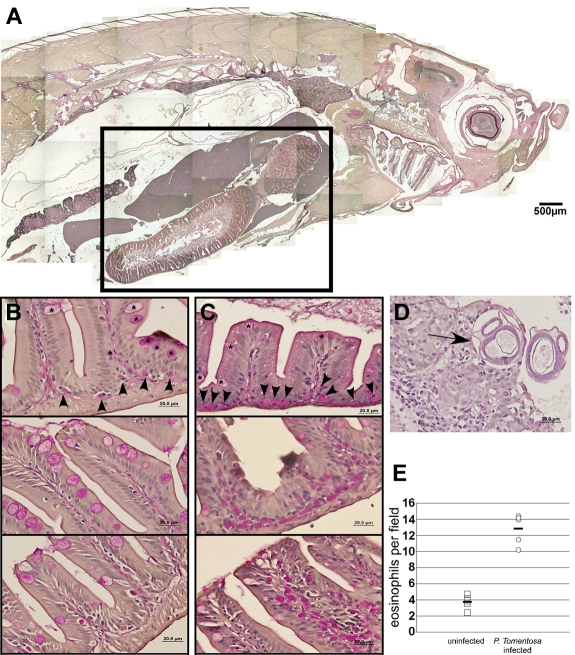
Eosinophils are granulocytic leukocytes implicated in numerous aspects of immunity and disease. The precise functions of eosinophils, however, remain enigmatic. Alternative models to study eosinophil biology may thus yield novel insights into their function. Eosinophilic cells have been observed in zebrafish but have not been thoroughly characterized. We used a gata2:eGFP transgenic animal to enable prospective isolation and characterization of zebrafish eosinophils, and demonstrate that all gata2(hi) cells in adult hematopoietic tissues are eosinophils. Although eosinophils are rare in most organs, they are readily isolated from whole kidney marrow and abundant within the peritoneal cavity. Molecular analyses demonstrate that zebrafish eosinophils express genes important for the activities of mammalian eosinophils. In addition, gata2(hi) cells degranulate in response to helminth extract. Chronic exposure to helminth- related allergens resulted in profound eosinophilia, demonstrating that eosinophil responses to allergens have been conserved over evolution. Importantly, infection of adult zebrafish with Pseudocapillaria tomentosa, a natural nematode pathogen of teleosts, caused marked increases in eosinophil number within the intestine. Together, these observations support a conserved role for eosinophils in the response to helminth antigens or infection and provide a new model to better understand how parasitic worms activate, co-opt, or evade the vertebrate immune response.


Figures

Figure 1
Figure 1
Analysis of hematopoietic cells from gata2eGFP transgenic fish by flow cytometry. Contour plots show the regional separation of major blood cell lineages by their light-scatter characteristics: FSCloSSCint-hi (R1, erythroid gate), FSCintSSClo (R2, lymphoid gate), FSChiSSClo (R3, precursor gate), FSChiSSCint (R4, myeloid gate), and FSChiSSChi (R5, eosinophil gate). Histograms indicate the abundance of GFP+ cells in each gate. (A) A representative scatter profile for WKM. Cells within each gate were quantified using the mean ± SD: R1, 37% ± 8.1%; R2, 17% ± 3.95%; R3, 6% ± 1.4%; R4, 24% ± 6.9%; and R5, 3% ± 1.3%. gata2hi cells were only present in the R5 gate: 74% ± 10%; n = 26. (B) A representative scatter profile for IPEX: R1, 9% ± 4.9%; R2, 10% ± 5.2%; R4', 7% ± 2.4%; and R5, 46% ± 18%. gata2hi cells were only present in the R5 gate: 91% ± 9.5%; n = 16.
Figure 2
Figure 2
gata2hi cells display the morphologic, histochemical, and ultrastructural properties of eosinophils. gata2hi and mpxhi cells were FACS sorted (purity ≥ 95%) from WKM (A-B) or IPEX (B). (A) Purified gata2hi (EOS) and mpxhi (NEU) cells were stained with hematoxylin and eosin, WG, PAS (red precipitate), MPX (brown precipitate), and TB (purple precipitate). Bar represents 5 μm. (B) Purified gata2hi cells were subjected to double staining with MPX and PAS or TEM analysis. A representation of each nuclear morphology (myelocyte, metamyelocyte, band, and polymorphonuclear) is shown as observed by cytospin (left) or TEM (right) analysis. Based on cytochemical analyses, the relative abundance (percentage) in either IPEX (top value) or WKM (bottom value) is provided for each morphologic class. Bar represents 2 μm. N indicates nucleus. Data are mean ± SD (n = 10).
Figure 3
Figure 3
gata2hi and mpxhi granulocytes differentially express genes that are important for eosinophil or neutrophil development and function, respectively. gata2hi and mpxhi cells were isolated by FACS (purity ≥ 95%) from WKM. Transcript abundance was measured from either gata2hi (black bars) or mpxhi cells (white bars) by quantitative polymerase chain reaction analysis. Expression of the gene of interest was normalized to that of ef1α. Data are mean ± SD (n = 3 or 4). *P < .05. **P < .001.
Figure 4
Figure 4
gata2hi eosinophils degranulate in response to H polygyrus helminth extracts. (A) IPEX from 3 WT fish was cultured in media containing either HpAg over a concentration gradient or in PBS alone for 4 hours. Cell-free supernatants were collected and assayed for peroxidase activity using the OPD assay. An average of the optical density (A492) for each dose was taken from 3 independent experiments. Error bars represent SD. (B) Time course of degranulation as measured by OPD assay. IPEX was cultured in media with 10 μg HpAg; then cell-free supernatants were assayed for degranulation. (C) Purified gata2hi cells (∼ 5 × 10⁴) from IPEX were lysed with 0.2% Triton-X or cultured for 2 hours in media with either 10 μg of HpAg or PBS. Cell-free supernatants were then collected and assayed for peroxidase activity using the OPD assay. Bars represent the mean optical density (A492) measured from 3 independent experiments with SD. *P < .05. **P < .001. (D) TEM photomicrographs show purified gata2hi cells (∼ 2.5 × 10⁵) cultured for 2 hours in media with either 12 μg of HpAg (iii-viii) or PBS (i-ii). Note heterogeneous populations of granules that display a range of electron lucency; granules are completely lucent (black arrowheads), marbled (white arrowheads), or variegated (white arrows). Electron-lucent granules were localized along cell peripheries (iii-vi). N indicates nucleus. Images are representative of 2 independent experiments.
Figure 5
Figure 5
Sustained exposure to papain or H polygyrus extract induces splenic and peripheral blood eosinophilia. (A) Spleens and (B) blood from treated and untreated adult gata2:eGFP+ zebrafish were analyzed for gata2hi leukocyte levels by FACS. Treated groups were injected weekly with papain or HpAg emulsified in IFA and tissues collected after one or 4 weeks. Control animals were either mock treated or uninjected. Bars represent means. *P < .05. **P < .001. Blood smears from fish immunized for 4 weeks with papain (C) or HpAg (D) showed elevated frequencies of PAS+ eosinophils. Images in panels C and D were acquired on an Olympus DP70 microscope with a 100× oil objective stained with PAS and hematoxylin. Images were collected with an Olympus DP Controller Version 2.1.1.183 and processed with Adobe Photoshop CS.
Figure 6
Figure 6
In vivo infection by P tomentosa leads to colonization of the gut and peritoneal cavity and an increase in local eosinophil number. (A) Composite image of sagittal section of adult zebrafish, stained with PAS. Inset: Area including intestines, shown in close-up views in panels B and C. (B) Imaging of the gut in uninfected animals shows nucleated PAS+ eosinophils interspersed along the basal lamina propria (arrowheads). Intestinal goblet cells (asterisks) are also PAS+, but distinct from eosinophils. (C) Infected animals show an increased number of PAS+ eosinophils along the basal lamina propria and also distally localized into villi. (D) P tomentosa larvae are observed within gut tissue (arrow). (E) Counts of eosinophils increase approximately 3-fold in intestines of infected animals. Images in panels A through D were acquired with an Olympus DP70 microscope and collected with Olympus DP Controller Version 2.1.1.183 and processed with Adobe Photoshop CS. Panel A is a composite image of a PAS and hematoxylin-stained section taken with a 10× dry objective. Images in panels B through D were taken with a 100× oil immersion objective and stained with PAS and hematoxylin.

Comment in

  • Eosinophils are in the swim!
    Rosenberg HF. Rosenberg HF. Blood. 2010 Nov 11;116(19):3692-3. doi: 10.1182/blood-2010-09-304345. Blood. 2010. PMID: 21071614 No abstract available.

Similar articles

Cited by

References

    1. Hogan SP, Rosenberg HF, Moqbel R, et al. Eosinophils: biological properties and role in health and disease. Clin Exp Allergy. 2008;38(5):709–750. - PubMed
    1. Rothenberg ME, Hogan SP. The eosinophil. Annu Rev Immunol. 2006;24:147–174. - PubMed
    1. Fabre V, Beiting DP, Bliss SK, et al. Eosinophil deficiency compromises parasite survival in chronic nematode infection. J Immunol. 2009;182(3):1577–1583. - PMC - PubMed
    1. Humbles AA, Lloyd CM, McMillan SJ, et al. A critical role for eosinophils in allergic airways remodeling. Science. 2004;305(5691):1776–1779. - PubMed
    1. Lee JJ, Dimina D, Macias MP, et al. Defining a link with asthma in mice congenitally deficient in eosinophils. Science. 2004;305(5691):1773–1776. - PubMed

Publication types

MeSH terms