. 2010 Sep;120(9):3255-66.
doi: 10.1172/JCI42957. Epub 2010 Aug 16.

Neurons derived from transplanted neural stem cells restore disrupted neuronal circuitry in a mouse model of spinal cord injury

Affiliations

Neurons derived from transplanted neural stem cells restore disrupted neuronal circuitry in a mouse model of spinal cord injury

Masahiko Abematsu et al. J Clin Invest. 2010 Sep.

Abstract

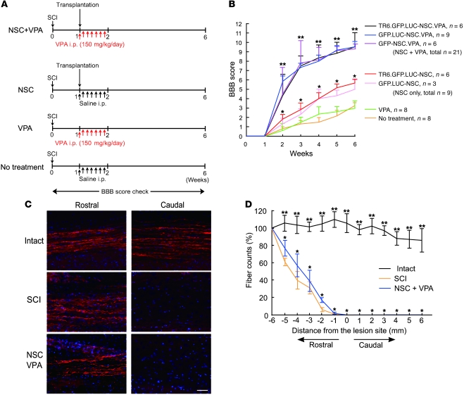
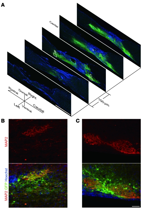
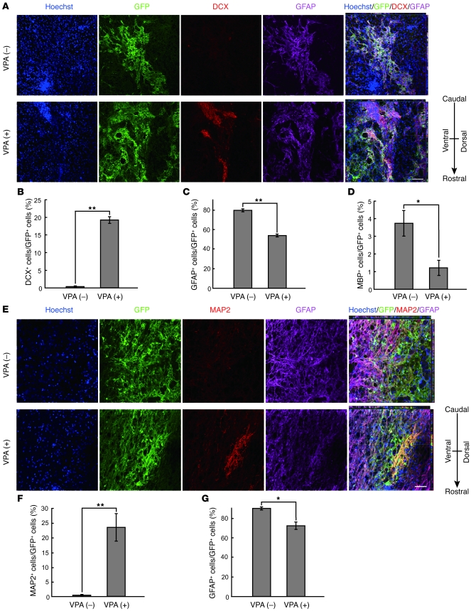
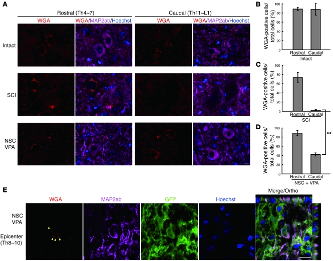
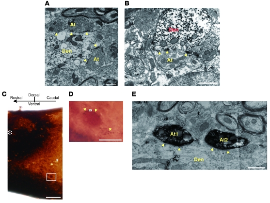
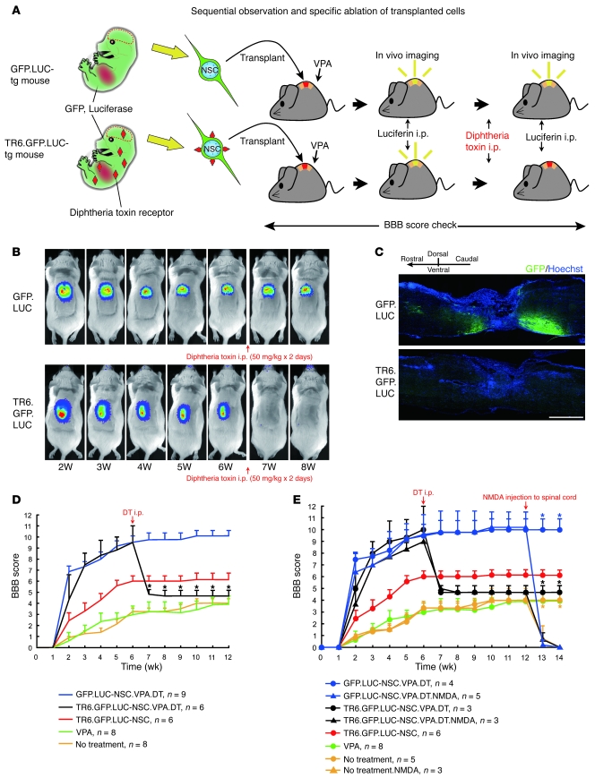
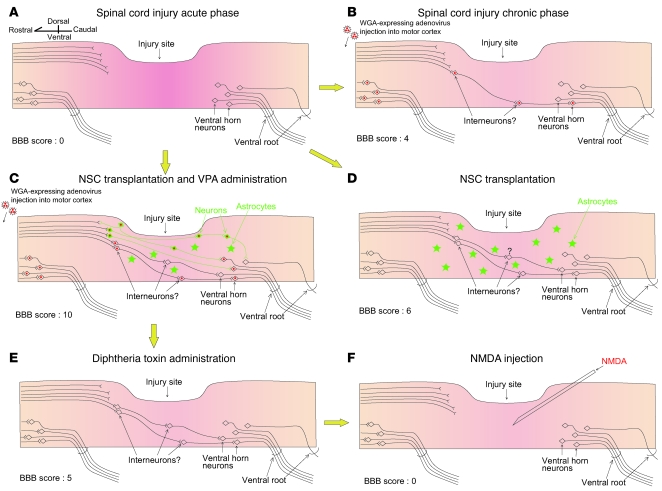
The body's capacity to restore damaged neural networks in the injured CNS is severely limited. Although various treatment regimens can partially alleviate spinal cord injury (SCI), the mechanisms responsible for symptomatic improvement remain elusive. Here, using a mouse model of SCI, we have shown that transplantation of neural stem cells (NSCs) together with administration of valproic acid (VPA), a known antiepileptic and histone deacetylase inhibitor, dramatically enhanced the restoration of hind limb function. VPA treatment promoted the differentiation of transplanted NSCs into neurons rather than glial cells. Transsynaptic anterograde corticospinal tract tracing revealed that transplant-derived neurons reconstructed broken neuronal circuits, and electron microscopic analysis revealed that the transplant-derived neurons both received and sent synaptic connections to endogenous neurons. Ablation of the transplanted cells abolished the recovery of hind limb motor function, confirming that NSC transplantation directly contributed to restored motor function. These findings raise the possibility that epigenetic status in transplanted NSCs can be manipulated to provide effective treatment for SCI.


Figures

Figure 1
Figure 1. A combination of NSC transplantation and VPA administration improves functional recovery of hind limbs without CST axon reextension.
(A) Schematic of the NSC transplantation and VPA injection protocol. (B) Time course of functional recovery of hind limbs after SCI. GFP-NSCs, GFP.LUC-NSCs, and TR6.GFP.LUC-NSCs were transplanted into the SCI epicenter 7 days after injury as indicated. Combined treatment with NSC transplantation and VPA administration resulted in the greatest functional recovery. Data represent mean ± SEM. **P < 0.001 compared with SCI models with no treatment; *P < 0.01 compared with SCI models with no treatment (repeated measures ANOVA). NSC+VPA, total n = 21. (C) Representative pictures of BDA-labeled CST fibers at 5 mm rostral and 5 mm caudal to the lesion site. BDA was injected into the motor cortices 12 weeks after SCI. 2 weeks after the injection, mice were fixed and spinal cord sections were stained. Representative results for a GFP-NSC–transplanted spinal cord are shown. Blue, Hoechst nuclear staining. Scale bar: 20 μm. (D) Quantification of the labeled CST fibers in the spinal cords of intact mice, SCI mice receiving no treatment, and SCI mice undergoing combined NSC/VPA treatment. Eight 30-μm–thick serial parasagittal sections from individual spinal cords were evaluated. The x axis indicates specific locations along the rostrocaudal axis of the spinal cord, and the y axis indicates the ratio of the number of BDA-labeled fibers at the indicated site to that at 6 mm rostral to the lesion site (Th9). **P < 0.001 compared with SCI models without treatment; *P = 0.188 There is no significant difference in the number of BDA-labeled fibers between NSC+VPA-treated mice (blue line) and SCI model mice with no treatment (yellow line) (repeated measures ANOVA). All data shown are from at least 3 experiments in parallel conditions, with error bars representing SEM.
Figure 2
Figure 2. Transplanted NSCs migrate from the injection site and encompass the lesion site.
Representative results of GFP-NSC–transplanted SCI model mice are shown. (A) A series of immunostaining images of injured spinal cord at 6 weeks after injury. SCI mice received combination treatment with NSC transplantation and VPA administration. Specimens were picked up every 150 μm and stained with anti-GFP (green) and MAP2 (not shown) antibodies and Hoechst (blue). The epicenter of the SCI is indicated (*). Scale bar: 1 mm. (B and C) Higher-magnification images of the white boxes in A. GFP-positive transplanted NSCs differentiated into MAP2-positive neurons and extended their processes. Scale bar: 50 μm.
Figure 3
Figure 3. VPA promotes neuronal differentiation of transplanted NSCs.
Representative results of GFP-NSC–transplanted SCI model mice are shown. (A) Confocal images of NSCs 1 week after transplantation into the injured spinal cords. Spinal cord sections from VPA-treated (+) and untreated (–) mice were stained with anti-GFP (green), anti-doublecortin (DCX) (immature neuronal marker, red) and anti-GFAP (magenta) antibodies, and Hoechst (blue). VPA administration resulted in an increase in the number of DCX-positive neuronal precursors among transplanted cells (lower panel). Scale bar: 20 μm. (BD) The percentages of DCX-, GFAP-, and MBP-positive cells in GFP-positive transplanted cells were quantified. **P < 0.01; *P < 0.05 compared with controls (Student’s t test). (E) Confocal images of NSCs 5 weeks after transplantation into injured spinal cords. Spinal cord sections from VPA-treated (+) and untreated (–) mice were stained with anti-GFP (green), anti-MAP2 (neuronal marker, red) and anti-GFAP (magenta) antibodies, and Hoechst (blue). VPA administration increased the numbers of MAP2-positive neurons (lower panel). Scale bar: 20 μm. (F and G) The percentages of cells positive for MAP2 or GFAP in GFP-positive transplanted cells in E were quantified. **P < 0.01; *P < 0.05 compared with control (Student’s t test). All data shown in BD, F, and G are from at least 15 confocal images of 3 individuals in parallel experiments, with error bars representing the SD.
Figure 4
Figure 4. Transplant-derived neurons reconstruct disrupted neuronal circuits in a relay manner.
(A) Representative pictures of WGA-labeled neuronal cell bodies located in the ventral horn at 14 weeks after SCI. Spinal cord sections were stained with anti-WGA (red) and -MAP2ab (magenta) antibodies and Hoechst (blue). Scale bar: 20 μm. Intense WGA immunoreactivity was observed as intracellular granule-like structures. Left panels show the rostral area (Th4–Th7), and right panels show the caudal area (Th11 to lumbar vertebra [L] 1). In uninjured mice, WGA injected into the bilateral motor cortices was transsynaptically transported to neurons in areas rostral and caudal to the injured site (top panels). In the SCI model mice that did not receive treatment, very little WGA was observed in caudal areas (middle panels). However, in spinal cords of animals that underwent dual treatment with NSC and VPA, WGA was clearly observed in neurons in the caudal areas (bottom panels). Representative results of GFP-NSC–transplanted SCI model mice are shown. (BD) The percentages of WGA-positive cells in the neurons localized in the ventral horn were quantified. **P < 0.05 (Student’s t test). All data shown are from at least 30 images, containing more than 600 cells, from 3 individuals (5 images per area) in parallel experiments, with error bars representing SD. (E) Representative confocal images of WGA-labeled transplant-derived MAP2-positive neurons. Sections were stained with anti-WGA (red), anti-MAP2ab (magenta) and anti-GFP (green) antibodies, and Hoechst (blue). Granule-like WGA structures (yellow arrowheads) could be seen in the GFP and MAP2ab–double-positive transplant-derived neurons. Scale bar: 10 μm.
Figure 5
Figure 5. Transplant-derived neurons make synapses with endogenous neurons.
(A) Immunoelectron microscopy image of a sagittal section of dual-treated (GFP-NSC and VPA) injured spinal cord (rostral area). A GFP-positive dendrite (Den) made synapses with GFP-negative endogenous axon termini (At) (yellow arrowheads). Scale bar: 1 μm. (B) In other rostral regions, a dendrite of a GFP-positive transplant-derived neuron made a synapse (yellow arrowheads) with the axon terminus of a GFP-negative endogenous neuron. Scale bar: 1 μm. (C) Sagittal section of dual-treated (NSC and VPA) injured spinal cord (caudal area) stained with anti-GFP antibody (dark brown). The epicenter of the SCI is indicated (*). Scale bar: 500 μm. (D) High-magnification image of a large neuron localized in the ventral horn in the white rectangle in C. GFP-positive transplanted neurons extended their processes toward an endogenous neuron (yellow arrowheads). Scale bar: 100 μm. (E) Immunoelectron microscopy image of the boxed area in D. GFP-positive axon termini made synapses with the dendrite of a GFP-negative endogenous large neuron (yellow arrowheads). Scale bar: 1 μm.
Figure 6
Figure 6. Ablation of transplanted cells abolishes hind limb motor function recovery.
(A) Schematic of the protocols for NSC transplantation and for detection and ablation of transplanted cells. NSCs derived from GFP.LUC- or TR6.GFP.LUC-Tg mice were transplanted into SCI model mice 1 week after injury. VPA was intraperitoneally administered every day for 1 week. Survival of transplanted cells and locomotor function of the mice were monitored weekly for 14 weeks. (B) Survival of transplanted cells was checked every week using a bioluminescence imaging system. 6 weeks after injury (5 weeks after transplantation), each mouse received 2 DT administrations. By the following week, LUC activity had completely disappeared in mice transplanted with TR6.GFP.LUC-NSCs (lower panel). (C) Sagittal sections from SCI model mice transplanted with GFP.LUC- and TR6.GFP.LUC-NSCs 2 weeks after DT injection. All transplanted cells were ablated with DT (lower panel). Scale bar: 1 mm. (D) Time course of the changes in BBB scores in SCI model mice. The hind limb function of mice that had undergone dual treatment with TR6.GFP.LUC-NSCs and VPA dropped drastically after DT administration (black line). *P < 0.0001 compared with GFP.LUC-NSC–transplanted, VPA-administered, and DT-injected SCI model mice (blue line) (repeated measures ANOVA). Data are mean ± SEM. VPA, n = 8; no treatment, n = 8. (E) Twelve weeks after injury, groups of SCI model mice received NMDA injections, as indicated, into the injury epicenter, to ablate local neurons in the gray matter (blue, black, and yellow lines with triangles). *P < 0.0001 compared with non-NMDA–injected mice in each group (blue, black, and yellow lines with circles) (repeated measures ANOVA). Data represent mean ± SEM.
Figure 7
Figure 7. Diagrammatic summary of destruction and reconstruction of neural circuits in the injured spinal cord.
(A) In the acute phase of SCI, CST fibers are disrupted and hind limb movement is completely lost. (B) In the chronic phase, WGA transport through the lesion site to the caudal area is negligible, probably because very few CST fibers are restored. (D) When NSCs were transplanted to the injured spinal cord, almost all of them differentiated into astrocytes, with the result that very little restoration of the injured CST occurred. (C) Following VPA administration, neuronal differentiation of transplanted NSCs was greatly enhanced and dramatic functional recovery could be observed. WGA is conveyed through the lesion site to the caudal area via transplant-derived neurons. (E) Ablation of transplant-derived cells with DT nullified hind limb functional recovery, suggesting that the transplanted cells contribute directly to the functional recovery in hind limbs. (F) Ablation of residual local neurons in the lesion site by NMDA led to a complete impairment of hind limb movement, suggesting that residual local endogenous neurons also play an important role in the improvement of hind limb motor function.

Comment in

Similar articles

Cited by

References

    1. Thuret S, Moon LD, Gage FH. Therapeutic interventions after spinal cord injury. Nat Rev Neurosci. 2006;7(8):628–643. doi: 10.1038/nrn1955. - DOI - PubMed
    1. David S, Aguayo AJ. Axonal elongation into peripheral nervous system “bridges” after central nervous system injury in adult rats. Science. 1981;214(4523):931–933. doi: 10.1126/science.6171034. - DOI - PubMed
    1. Richardson PM, McGuinness UM, Aguayo AJ. Axons from CNS neurons regenerate into PNS grafts. Nature. 1980;284(5753):264–265. doi: 10.1038/284264a0. - DOI - PubMed
    1. Schnell L, Schwab ME. Axonal regeneration in the rat spinal cord produced by an antibody against myelin-associated neurite growth inhibitors. Nature. 1990;343(6255):269–272. doi: 10.1038/343269a0. - DOI - PubMed
    1. Bregman BS, Kunkel-Bagden E, Schnell L, Dai HN, Gao D, Schwab ME. Recovery from spinal cord injury mediated by antibodies to neurite growth inhibitors. Nature. 1995;378(6556):498–501. doi: 10.1038/378498a0. - DOI - PubMed

Publication types

Substances