. 2010;92(4):224-34.
doi: 10.1159/000319257. Epub 2010 Aug 12.

Interactions of corticotropin-releasing factor, urocortin and citalopram in a primate model of stress-induced amenorrhea

Affiliations

Interactions of corticotropin-releasing factor, urocortin and citalopram in a primate model of stress-induced amenorrhea

Karin V Weissheimer et al. Neuroendocrinology. 2010.

Abstract

Background/aims: We established a cynomolgus macaque model of stress-induced amenorrhea in which the application of combined metabolic and psychosocial stress suppressed ovulation in stress-sensitive (SS) individuals, but not in highly stress-resilient (HSR) individuals. We previously reported that SS monkeys have deficits in global serotonin release and serotonin-related gene expression in the raphe nucleus, and that administration of the selective serotonin reuptake inhibitor S-citalopram increased estrogen and progesterone production in SS monkeys. In this study, we questioned whether there was a difference in corticotropin-releasing factor (CRF) or urocortin (UCN) stress-related peptide systems in the midbrain raphe region when HSR and SS monkeys treated with placebo or S-citalopram are compared.

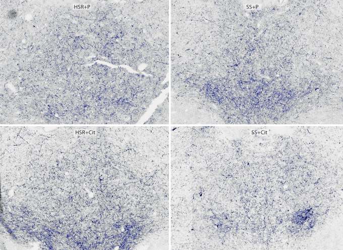
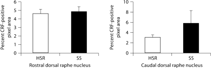
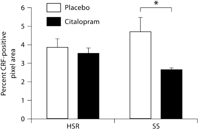
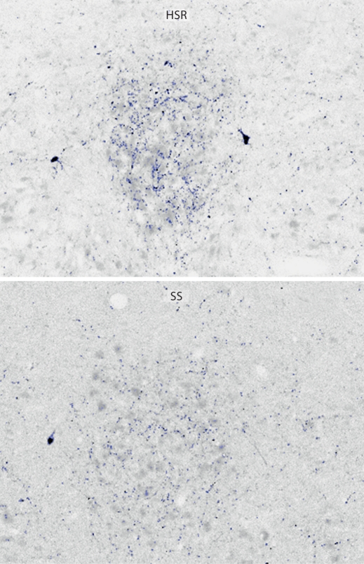
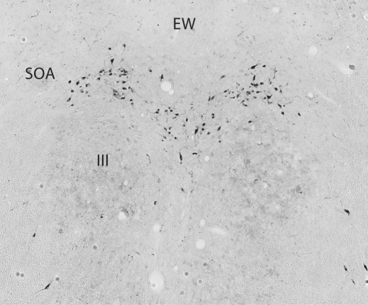
Methods: Monkeys characterized as HSR or SS were administered placebo or S-citalopram for 15 weeks. CRF fibers in the dorsal raphe were detected with an antibody against human CRF. UCN1 fibers were immunostained in an area rostral to the dorsal raphe. The fibers were quantified by stereology and analyzed by two-way ANOVA. UCN1 cell bodies were counted in the supraoculomotor area near the Edinger-Westphal nucleus.

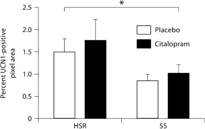
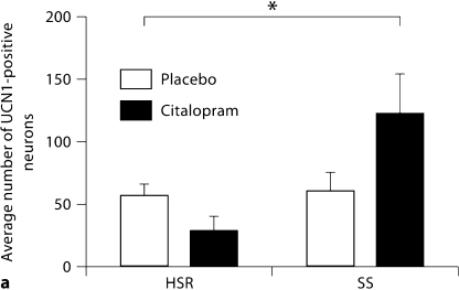
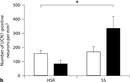
Results: S-citalopram significantly decreased the CRF fiber density in SS animals, but not in HSR animals. SS monkeys had a significantly lower UCN1 fiber density compared to HSR monkeys, but S-citalopram treatment did not alter the UCN1 fiber density. SS animals treated with S-citalopram tended to have a higher number of UCN1-positive cell bodies than the other groups.

Conclusion: S-citalopram decreased CRF fiber density and appears to increase the production of UCN1 in SS individuals, indicating the likelihood that serotonin is involved in regulating CRF and UCN1 in individuals who are sensitive to the effects of serotonin.


Figures

Fig. 1
Fig. 1
Photomicrographs of CRF fiber staining in the dorsal raphe nucleus. Stereology montage of CRF fiber staining in the dorsal raphe nucleus in a representative animal from each treatment group. Sections were immunostained for CRF and segmented into positively and negatively stained pixels. Blue: positive pixels. Cell bodies or debris were erased prior to pixel quantitation. Visually, there appears to be a lower density of CRF fibers in the SS+Cit animal. ×10.
Fig. 2
Fig. 2
CRF fiber density expressed as percent of total pixel area covered by CRF-positive fibers (pixels) in the Pb-treated groups. Rostral and caudal regions of the dorsal raphe nucleus of the HSR and SS groups treated with placebo (HSR+Pb and SS+Pb, respectively). In the rostral region, there was no significant difference between the groups. In caudal region, there was a significant difference (F test: p = 0.0170) between HSR+Pb and SS+Pb groups, with the SS+Pb group exhibiting a significantly higher CRF fiber density compared to the HSR+Pb group.
Fig. 3
Fig. 3
Average CRF-positive pixels across 4 levels of the dorsal raphe nucleus in all groups expressed as percent of total area examined. There was a significant effect of treatment (p = 0.0496), but no effect of stress sensitivity (p = 0.6755) and no interaction (p = 0.3027). The SS+Cit group exhibited a significantly lower CRF fiber density compared to the SS+Pb group. * Significant difference (Bonferroni: p < 0.05).
Fig. 4
Fig. 4
Photomicrographs of UCN1-positive fibers in the midbrain, rostral to the dorsal raphe nucleus, in a representative HSR animal and an SS animal. Sections were immunostained for UCN1 and segmented into positively and negatively stained pixels. Blue: positive pixels. Cell bodies or debris were erased prior to pixel quantitation. Visually, there were few UCN1 fibers in the SS groups.
Fig. 5
Fig. 5
Average UCN1-positive pixels across 3 levels of the rostral midbrain in all groups expressed as percent of total area examined. There was a significant effect of stress sensitivity (p = 0.0477), but no effect of S-citalopram treatment (p = 0.4958) and no interaction (p = 0.8946). The SS groups exhibited a significantly lower UCN1 fiber density compared to the HSR groups. * Significant difference (ANOVA: p < 0.05).
Fig. 6
Fig. 6
Photomicrograph of UCN1-positive cells in the SOA near the Edinger-Westphal nucleus (EW) in a representative animal. The cells are superior to the oculomotor (third) nucleus (III).
Fig. 7
Fig. 7
Overall averages of UCN1-positive cells in the SOA. The number of neurons across 4 levels was summed, generating the total number of neurons per animal. Then, the average of the animal totals was obtained for each group. * Significant difference (ANOVA: p < 0.05). a Average number of UCN1-positive neurons. By two-way ANOVA, there was a significant effect of stress sensitivity (p = 0.04), but no effect of treatment (p = 0.43). There was a nearly significant interaction (p = 0.056), indicating that S-citalopram increased detectable UCN1 neurons in the SS group, but not in the HSR group. b Average number of UCN1-positive neurons per cubic millimeter for each group. By two-way ANOVA, there was a significant effect of stress sensitivity (p = 0.04), but no effect of treatment (p = 0.43). There was a nearly significant interaction (p = 0.056), indicating that S-citalopram increased detectable UCN1 neurons in the SS group, but not in the HSR group.
Fig. 7
Fig. 7
Overall averages of UCN1-positive cells in the SOA. The number of neurons across 4 levels was summed, generating the total number of neurons per animal. Then, the average of the animal totals was obtained for each group. * Significant difference (ANOVA: p < 0.05). a Average number of UCN1-positive neurons. By two-way ANOVA, there was a significant effect of stress sensitivity (p = 0.04), but no effect of treatment (p = 0.43). There was a nearly significant interaction (p = 0.056), indicating that S-citalopram increased detectable UCN1 neurons in the SS group, but not in the HSR group. b Average number of UCN1-positive neurons per cubic millimeter for each group. By two-way ANOVA, there was a significant effect of stress sensitivity (p = 0.04), but no effect of treatment (p = 0.43). There was a nearly significant interaction (p = 0.056), indicating that S-citalopram increased detectable UCN1 neurons in the SS group, but not in the HSR group.

Similar articles

Cited by

References

    1. Boivin J, Bunting L, Collins JA, Nygren KG. International estimates of infertility prevalence and treatment-seeking: potential need and demand for infertility medical care. Hum Reprod. 2007;22:1506–1512. - PubMed
    1. Reindollar RH, Novak M, Tho SP, McDonough PG. Adult-onset amenorrhea: a study of 262 patients. Am J Obstet Gynecol. 1986;155:531–543. - PubMed
    1. Berga SL, Loucks TL. Use of cognitive behavior therapy for functional hypothalamic amenorrhea. Ann NY Acad Sci. 2006;1092:114–129. - PubMed
    1. Marcus MD, Loucks TL, Berga SL. Psychological correlates of functional hypothalamic amenorrhea. Fertil Steril. 2001;76:310–316. - PubMed
    1. Williams NI, Berga SL, Cameron JL. Synergism between psychosocial and metabolic stressors: impact on reproductive function in cynomolgus monkeys. Am J Physiol Endocrinol Metab. 2007;293:E270–E276. - PubMed

Publication types

MeSH terms