. 2011 Feb;21(2):275-89.
doi: 10.1038/cr.2010.118. Epub 2010 Aug 17.

The TWIST/Mi2/NuRD protein complex and its essential role in cancer metastasis

Affiliations

The TWIST/Mi2/NuRD protein complex and its essential role in cancer metastasis

Junjiang Fu et al. Cell Res. 2011 Feb.

Abstract

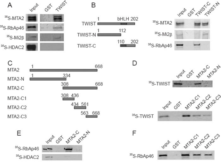
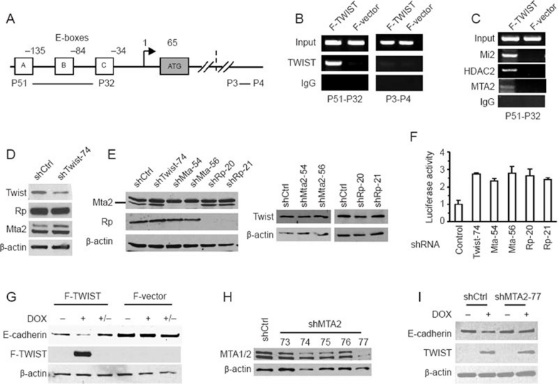
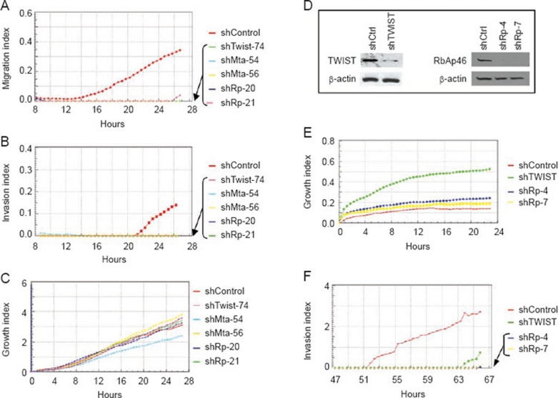
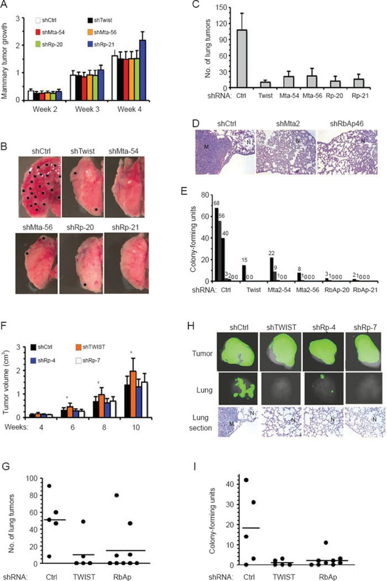
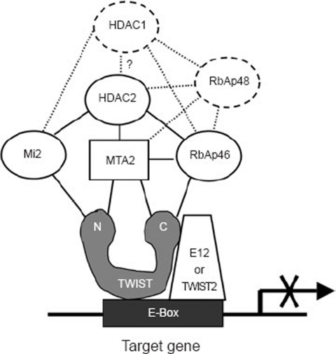
The epithelial-mesenchymal transition (EMT) converts epithelial tumor cells into invasive and metastatic cancer cells, leading to mortality in cancer patients. Although TWIST is a master regulator of EMT and metastasis for breast and other cancers, the mechanisms responsible for TWIST-mediated gene transcription remain unknown. In this study, purification and characterization of the TWIST protein complex revealed that TWIST interacts with several components of the Mi2/nucleosome remodeling and deacetylase (Mi2/NuRD) complex, MTA2, RbAp46, Mi2 and HDAC2, and recruits them to the proximal regions of the E-cadherin promoter for transcriptional repression. Depletion of these TWIST complex components from cancer cell lines that depend on TWIST for metastasis efficiently suppresses cell migration and invasion in culture and lung metastasis in mice. These findings not only provide novel mechanistic and functional links between TWIST and the Mi2/NuRD complex but also establish new essential roles for the components of Mi2/NuRD complex in cancer metastasis.


Figures

Figure 1
Figure 1
Purification and characterization of TWIST interacting proteins. (A) Induction of Flag-tagged TWIST (F-TWIST) protein. Stable HEK293 cells with F-TWIST or control F-vector were treated with vehicle or doxycyclin (DOX) for 6 h. Western blot analysis was performed with whole-cell extracts and antibodies against Flag and β-actin. (B) Purification of TWIST protein complex. Flag antibody beads were incubated with F-TWIST and F-vector cell lysates. The immunopurified protein complexes were resolved by 4-20% SDS-PAGE and visualized by Imperial Protein Stain. Mass spectrometry analysis identified MTA2 and E12 proteins from the gel slice a (GS-a), HDAC2 and RbAp46 proteins from GS-b and TWIST1 and TWIST2 from GS-c of the F-TWIST gel lane. ns, non-specific bands appearing in both lanes. (C) The cell lysate was prepared in cell lysis buffer with 0.5% of NP-40 from 293 cells. The control (Ctrl) cell lysate was prepared with sonication for extracting genomic DNA as a positive control. PCR was performed to detect E-cadherin promoter in both cell lysates. (D) Co-IP and western blot. Endogenous TWIST protein in MDA-MB-435 cells was immunoprecipitated with TWIST antibody and control IgG. Associated proteins were assayed with indicated antibodies. (E) Detection of HDAC activity in TWIST protein complex. Chemical assay for HDAC activity was performed with [H3]-acetylated-histone H4 peptide as a substrate and co-immunoprecipitated materials by Flag antibody from F-TWIST and F-vector cells. The reactions were carried out with or without HDAC inhibitor sodium butyrate.
Figure 2
Figure 2
TWIST directly interacts with multiple components of the Mi2/NuRD/MTA2 complex. (A) GST pull-down assays. GST-TWIST and GST proteins purified from bacteria were mixed with 35S-labeled MTA2, Mi2, RbAp46 and HDAC2 proteins produced from transcription and translation-coupled reactions. The 35S-labeled proteins bound to GST and GST-TWIST were resolved by SDS-PAGE and visualized by exposing to X-ray films. (B) Full-length TWIST and its N- and C-terminal fragments used in GST-fusion proteins and GST pull-down assays using GST, GST-TWIST-N and GST-TWIST-C and 35S-labeled MTA2, Mi2 and RbAP46 as indicated. (C) Full-length MTA2 and its fragments used in GST-fusion proteins. (D) GST pull-down assays using GST-fusion proteins containing MTA2 and its fragments and 35S-labeled TWIST. (E, F) GST pull-down assays using GST-fusion proteins containing MTA2 and its fragments and 35S-labeled RbAp46 and HDAC2.
Figure 3
Figure 3
TWIST recruits the Mi2/NuRD/MTA2 complex to E-cadherin promoter and depends on MTA2 to repress E-cadherin expression. (A) The proximal region of the E-cadherin promoter contains three TWIST-binding E-boxes. In the ChIP assay, PCR with P51 and P32 primers was used to amplify the region with these E-boxes. PCR with primers P3 and P4 in the 3′ region of the E-cadherin gene, which was more than 100 kb downstream from the indicated E-boxes, served as negative control in the ChIP assay. (B) ChIP assays using F-TWIST and F-vector HEK293 cells, Flag antibody or control IgG and primer pairs of P51/P32 and P3/P4 for E-cadherin gene. (C) ChIP-re-ChIP assays for the E-box region of the E-cadherin gene. The first-step immunoprecipitation was performed with Flag antibody and cross-linked and sonicated chromatin of F-TWIST and F-vector HEK293 cells. The second-step immunoprecipitation was performed with the eluted materials from the first step and with antibodies against Mi2, HDAC2 and MTA2 as indicated. IgG served as a negative control in the second step ChIP. (D, E) Generation of stable 4T1 cell pools with lentivirus-mediated expression of Twist shRNA (shTwist-74), Mta2 shRNA (shMta-54 and -56), RbAp46 shRNA (shRp-20 and -21) and non-targeting shRNA (shCtrl). Panel D shows efficient knockdown of Twist and the knockdown did not affect RbAp46 (Rp) and Mta2. Panel E shows efficient knockdown of Mta2 and RbAp46 proteins, and knockdown of any one of two proteins did not affect the other one (left panel) and also did not affect Twist (right panel). (F) Knockdown of Twist, Mta2 or RbAp46 in 4T1 cells increases E-cadherin promoter activity. 4T1 cell pools carrying non-targeting shRNA (control) and targeting shRNAs for Twist, Mta2 and RbAp46 as indicated were transfected with 100 ng of the E-Cad-promoter-Luciferase reporter plasmid and 30 ng of β-gal expression plasmids in 24-well plates. Luciferase activity was assayed 40 h later, normalized to β-gal activity and presented as mean ± SD, n = 3. (G) TWIST represses endogenous E-cadherin expression. HEK293 cells with F-TWIST and F-vector were set up in three groups. Groups 1 and 2 were treated with vehicle (lane 1 and 4) and DOX for 2 days to induce F-TWIST (lanes 2 and 3) and Flag (lanes 5 and 6). Group 3 was treated with DOX for 2 days, and then DOX was removed and cells were cultured for 7 more days (lanes 3 and 6). Western blot analyses were performed to analyze E-cadherin, F-TWIST and β-actin. (H) Western blot analysis of MTA2 in puromycin-selected stable F-TWIST HEK293 cells infected by lentiviruses with a non-targeting control shRNA and five different shRNAs against human MTA2 mRNA. The shRNA-MTA2-77 lentivirus successfully reduced both MTA2 and MTA1 proteins. (I) MTA2 knockdown blocks TWIST-mediated suppression of E-cadherin expression. shRNA control and shRNA-MTA2-77 cells were treated with vehicle or DOX to induce F-TWIST for 3 days before western blot was performed to assay E-cadherin, F-TWIST and β-actin.
Figure 4
Figure 4
Knockdown of TWIST, MTA2 or RbAp46 specifically inhibits cancer cell migration and invasion. (A-C) Real-time assays of cell migration, invasion and growth for 4T1 cells with non-targeting shRNA (shControl) and targeting shRNAs specific to Twist (shTwist-74), Mta2 (shMta-54 and shMta-56) and RbAp46 (shRp-20 and shRp-21). (D) Western blot analysis for knockdown of TWIST and RbAp46 in MDA-MB-435 cells by shRNAs. (E, F) Real-time assays of cell growth and invasion for MDA-MB-435 cell pools expressing shControl, shTWIST and shRbAp46 (shRp-4 and shRp-7).
Figure 5
Figure 5
Twist, Mta2 and RbAp46 are required for breast cancer cell metastasis in mice. (A) Growth profiles of the isografted mammary tumors from the indicated 4T1 cells in female BALB/c recipient mice. Data at the 2nd and 3rd weeks are presented as estimated average tumor volumes (cm3). Data at the 4th week are presented as average tumor weight (g). (B) Representative photos of the lungs from different recipient mice carrying isografted mammary tumors from the indicated 4T1 cells at the 4th week. The asterisks indicate large metastatic tumors. (C) Average numbers of visible 4T1 metastatic nodules in individual mouse lung were counted and are presented as mean ± SD, n = 5. (D) Representative H&E-stained lung sections prepared from recipient mice bearing isografted mammary tumors formed from shControl, shMta2 and shRbAp46 4T1 cells. N, normal lung tissue; M, metastatic nodule. (E) Number of 4T1 tumor cell colonies formed in 15-day culture using 150 μl of blood collected from recipient mice bearing the indicated types of 4T1 tumors for 4 weeks. (F) MDA-MB-435 xenograft tumor growth in athymic mice. MDA-MB-435 cells expressing shControl (shCtrl), shTWIST and shRbAp46 (shRp-4 and shRp-7) were injected into the mammary fat pads and tumor volumes were measured at the indicated time points. Ten tumors in each of the shControl, shTWIST and shRp-4 groups and eight tumors in the shRp-7 group were measured. (G) Representative tGFP images of primary tumors and lung metastasis derived from MDA-MB-435 cells with shControl, shTWIST and shRbAp46. H&E-stained lung sections prepared from mice with the indicated types of tumors are also presented. M, metastatic nodule; N, normal lung tissue. (H) Number of MDA-MB-435 lung tumors observed under a stereomicroscope. The average tumor number of the shControl group (n = 5) is significantly higher than that observed in the shTWIST (n = 5) and shRbAp46 (n = 9) groups (P < 0.05). (I) Number of tumor cell colonies formed in a culture containing 150 μl of blood collected from recipient mice with the indicated types of MDA-MB-435 tumors. The average number of colonies formed from the blood of mice with shControl tumors is significantly higher than that formed from the blood samples of mice with shTWIST and shRbAp46 tumors (P < 0.05).
Figure 6
Figure 6
A model for TWIST-mediated gene repression. TWIST binds to E-box as homodimer or heterodimer with E12 or with TWIST2. The TWIST N-terminus interacts with MTA2 and Mi2, while its C-terminus interacts with MTA2 and RbAp46. Mi2, RbAp46 and MTA2 recruit HDAC2 to the DNA/TWIST complex through direct interactions. The TWIST-dependent recruitment of Mi2 and HDAC2 chromatin-remodeling enzymes to the promoter represses the target gene expression.

References

    1. Weinberg RA.The Biology of CancerNew York: Garland Science, 2007
    1. Onder TT, Gupta PB, Mani SA, et al. Loss of E-cadherin promotes metastasis via multiple downstream transcriptional pathways. Cancer Res. 2008;68:3645–3654. - PubMed
    1. Yang J, Mani SA, Donaher JL, et al. Twist, a master regulator of morphogenesis, plays an essential role in tumor metastasis. Cell. 2004;117:927–939. - PubMed
    1. Yang J, Mani SA, Weinberg RA. Exploring a new twist on tumor metastasis. Cancer Res. 2006;66:4549–4552. - PubMed
    1. Castanon I, Baylies MK. A Twist in fate: evolutionary comparison of Twist structure and function. Gene. 2002;287:11–22. - PubMed

Publication types

MeSH terms