. 2010 Aug 12;6(8):e1001041.
doi: 10.1371/journal.ppat.1001041.

The human fungal pathogen Cryptococcus neoformans escapes macrophages by a phagosome emptying mechanism that is inhibited by Arp2/3 complex-mediated actin polymerisation

Affiliations

The human fungal pathogen Cryptococcus neoformans escapes macrophages by a phagosome emptying mechanism that is inhibited by Arp2/3 complex-mediated actin polymerisation

Simon A Johnston et al. PLoS Pathog. .

Abstract

The lysis of infected cells by disease-causing microorganisms is an efficient but risky strategy for disseminated infection, as it exposes the pathogen to the full repertoire of the host's immune system. Cryptococcus neoformans is a widespread fungal pathogen that causes a fatal meningitis in HIV and other immunocompromised patients. Following intracellular growth, cryptococci are able to escape their host cells by a non-lytic expulsive mechanism that may contribute to the invasion of the central nervous system. Non-lytic escape is also exhibited by some bacterial pathogens and is likely to facilitate long-term avoidance of the host immune system during latency. Here we show that phagosomes containing intracellular cryptococci undergo repeated cycles of actin polymerisation. These actin 'flashes' occur in both murine and human macrophages and are dependent on classical WASP-Arp2/3 complex mediated actin filament nucleation. Three dimensional confocal imaging time lapse revealed that such flashes are highly dynamic actin cages that form around the phagosome. Using fluorescent dextran as a phagosome membrane integrity probe, we find that the non-lytic expulsion of Cryptococcus occurs through fusion of the phagosome and plasma membranes and that, prior to expulsion, 95% of phagosomes become permeabilised, an event that is immediately followed by an actin flash. By using pharmacological agents to modulate both actin dynamics and upstream signalling events, we show that flash occurrence is inversely related to cryptococcal expulsion, suggesting that flashes may act to temporarily inhibit expulsion from infected phagocytes. In conclusion, our data reveal the existence of a novel actin-dependent process on phagosomes containing cryptococci that acts as a potential block to expulsion of Cryptococcus and may have significant implications for the dissemination of, and CNS invasion by, this organism.


Conflict of interest statement

One of the authors (RCM) is an editor for PLoS Pathogens and PLoS ONE. We declare no financial interest in this manuscript.

Figures

Figure 1
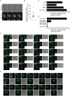
Figure 1. A dynamic actin coat transiently and repeatedly assembles around Cryptococcus-containing phagosomes.
(A) Brightfield and actin-GFP epifluorescence images of RAW macrophage-like cell line stably expressing actin-GFP. Actin is transiently recruited to the phagosome (120s and 240s) containing a phagocytosed Cryptococcus. (B) Box plot of the number of flashes that occur on individual phagosomes. Brightfield and actin-GFP epifluorescence images of RAW macrophage-like cells stably expressing actin-GFP were captured every 2 minutes for 18 hours post phagocytosis of Cryptococcus and scored for actin flashes. (45 phagosomes, n = 5 experiments; excludes phagosomes that do not flash; for explanation of box dimensions see materials and methods). (C) The proportion of Cryptococcus containing phagosomes that exhibit at least one actin flash over 18 hours. Actin flashes occur on phagosomes of both serotypes of Cryptococcus neoformans. Serotype D: JEC21 n = 2; B3501 n = 2; Serotype A: ATCC90112 n = 6; H99 n = 3; (D) Comparison of the incidence of actin flashing on ATCC90112 containing phagosomes (reproduced from (C)) to heat killed ATCC90112 (n = 10, P = 2.3×10⁻⁹), latex beads (n = 12, P = 1.1×10⁻³²) and IgG coated latex beads (n = 3 P = 8.7×10⁻³⁰). Statistical significance is indicated by an asterisk and was tested with comparison to ATCC90112 using Fisher's exact test. (E) Three dimensional time lapse confocal of an actin flash around a Cryptococcus containing phagosome in RAW macrophage-like cell stably expressing actin-GFP. Images are representative of >100 cells observed over 5 independent experiments. (F) Three dimensional time lapse confocal of an actin flash around a Cryptococcus containing phagosome in human primary macrophage transformed with Lifeact actin sensor peptide tagged with GFP. Red arrow indicates the location of the phagosome in transmitted light images (note that it is obscured in some panels due to the large number of cortical membrane ruffles in these cells). Images are representative of 30 cells observed over 3 independent experiments. TL, transmitted light. All scale bars 10µm.
Figure 2
Figure 2. Actin flashes are generated by the Arp2/3-WASP pathway.
(A) Transmitted and three dimensional confocal (A1R) fluorescence image of actin flash in J774 macrophage-like cells. Endogenous actin was labelled with filamentous actin binding drug phalloidin after fixation, 3 hours post phagocytosis. Scale bar 10µm. Image is representative of >100 cells observed over 3 independent experiments. (B) Transmitted and three dimensional confocal (A1R) fluorescence image of actin flash in human primary macrophages. Endogenous actin was labelled with filamentous actin binding drug phalloidin after fixation, 3 hours post phagocytosis. Scale bar 10µm. Image is representative of >100 cells observed over 3 independent experiments. (C) Three dimensional confocal (SP2) fluorescence image of a representative flashing J774 macrophage labelled for F-actin and Arp2/3 complex member ARPC2 after fixation, 3 hours post phagocytosis. Actin and Arp2/3 complex are both present on flashing phagosomes. Scale bar 5 µm. (D) Co-localisation of ARPC2 and F-actin, co-localised pixels are shown in white and determined as described in materials and methods. Actin and Arp2/3 complex show a high degree of co-localisation on phagosomes containing Cryptococcus. Scale bar 5 µm. (E) Proportion of phagosomes that flash in J774 macrophages following inhibition of WASP/N-WASP by wiskostatin. Cells were incubated with the indicated concentrations of wiskostatin for three hours post phagocytosis before fixation and labelling of filamentous actin. P-values are: 1nM = 0.025, 10nM = 0.011, 100nM = 2.0×10⁻⁴ and 1 µm = 1.3×10⁻⁵. Statistical significance tested with comparison to untreated using Fisher's exact test, >1000 phagosomes were scored for each concentration from a total of n = 4 experiments.
Figure 3
Figure 3. Actin flashes are positively correlated with cryptococcal expulsion but do not contribute to force generation.
(A) Total proportion of phagosome lifetime for which an actin flash is present, comparing retained cryptococci versus those subsequently expelled. The phagosomes of expelled cryptococci spend twice the proportion of their lifetime undergoing actin flashing (mean = 0.32, median = 0.27) as those that are retained (mean = 0.16, median  = 0.08) (B) Four Cryptococcus strains were scored for the incidence of expulsion and flashing. Strains were selected to give a wide range of expulsion incidence; expulsion incidence had been previously identified for large number of strains during preliminary experiments. Both scores were performed simultaneously on the same cells. (C) Three dimensional time lapse confocal of the expulsion of Cryptococcus from RAW macrophage-like cells stably expressing actin-GFP. Red line shows the position of the phagosome in the z-axis. x,y image is the confocal z-axis slice corresponding to the position of the red line (i.e. through the centre of the phagosome). The cryptococcal phagosome is stably positioned within the cell (0–60 minutes). Two minutes prior to expulsion the phagosome moves upwards, at 60+90s can be seen partially ejected and at 60+120s has completely exited the cell. Immediately following expulsion the cryptococcal cell moves away from the macrophage. There is no increase in actin fluorescence during expulsion. Scale bar is 10 µm.
Figure 4
Figure 4. Modulation of actin flashes influences the incidence of expulsion.
Actin flashes were modulated by inhibiting actin depolymerisation with 50 nM jasplakinolide, actin polymerisation with 10nM cytochalasin or WASP/N-WASP (and therefore Arp2/3 complex mediated actin polymerisation) with 100nM wiskostatin. (A) Percentage of phagosomes that flash in J774 macrophages with modulation of actin. Jasplakinolide, P = 1.1×10⁻⁴; cytochalasin D, P = 4.2×10⁻⁸; wiskostatin, P = 5.0×10⁻¹⁰. (B) Percentage of phagosomes that flash in human primary macrophages with modulation of actin. Jasplakinolide, P = 6.7×10⁻⁵; cytochalasin D, P = 2.9×10⁻⁸; wiskostatin, P = 3.4×10⁻⁶. (C) Box plots of actin flash length in RAW macrophage-like cells stably expressing actin-GFP. Horizontal red line indicates the mean. Stabilisation of actin filaments with jasplakinolide lengthens flashes while destabilising actin filaments with cytochalasin D shortens flashes. Note that the lower threshold of flash length is constrained by the frame rate of the time lapse imaging. Untreated, 68 phagosomes, n = 5 experiments; jasplakinolide, 138 phagosomes, n = 9 experiments; cytochalasin, 77 phagosomes, n = 9 experiments (D) Modulation of expulsion of cryptococci from RAW macrophage-like cells stably expressing actin-GFP. Expulsion of cryptococci is depicted as the proportion of phagosomes ‘at risk’ (i.e. remaining intracellular), calculated every 600s for each treatment. Influence of actin drug treatments on expulsion is the inverse of that seen with flash length. Untreated, 68 phagosomes, n = 5 experiments; jasplakinolide, 138 phagosomes, n = 9 experiments; cytochalasin, 77 phagosomes, n = 9 experiments; wiskostatin, 66 phagosomes, n = 4 experiments. (E) Modulation of expulsion of cryptococci from human primary macrophages. Expulsion of cryptococci depicted as the proportion of phagosomes ‘at risk’, calculated every 600s for each treatment. As with J774 macrophages, the influence of actin drug treatments on expulsion in human primary macrophages is the inverse of that seen with flash length. Untreated, 75 phagosomes, n = 3 experiments; jasplakinolide, 51 phagosomes, n = 4 experiments; cytochalasin, 27 phagosomes, n = 6 experiments; wiskostatin, 24 phagosomes, n = 3 experiments.
Figure 5
Figure 5. Actin flashes immediately follow rapid permeabilisation of phagosomes containing Cryptococcus.
(A) J774 macrophages that had phagocytosed either IgG coated latex beads or Cryptococcus in the presence of FITC dextran (see figure S6) were fixed at 0 and 3 hours post phagocytosis (hpp) and scored for the presence of dextran (latex beads 0h = 450 phagosomes, 3h = 177 phagosomes; ATCC90112 0h = 1287 phagosomes, 3h = 1670 phagosomes from 3 independent experiments). (B, C) Dextran-positive phagosomes were never observed with actin flashes (B) and vice versa (C). Image panels are single slices from a confocal (SP2) z-stack. Transmitted light channel is labelled as TL. Scale bars 5 µm. (D) Time lapse phase contrast and epifluorescence images of a RAW macrophage-like cell stably expressing actin-GFP with TRITC dextran labelled, Cryptococcus-containing phagosomes. Phagosome permeabilisation is indicated by the loss of TRITC dextran (48:35–50:35). Note that the loss of dextran is immediately followed by an actin flash. Scale bar 5 µm.

References

    1. Freitag NE, Port GC, Miner MD. Listeria monocytogenes - from saprophyte to intracellular pathogen. Nat Rev Microbiol. 2009;7:623–628. - PMC - PubMed
    1. Pieters J. Mycobacterium tuberculosis and the macrophage: maintaining a balance. Cell Host Microbe. 2008;3:399–407. - PubMed
    1. Prost LR, Sanowar S, Miller SI. Salmonella sensing of anti-microbial mechanisms to promote survival within macrophages. Immunol Rev. 2007;219:55–65. - PubMed
    1. Alvarez M, Casadevall A. Phagosome extrusion and host-cell survival after Cryptococcus neoformans phagocytosis by macrophages. Curr Biol. 2006;16:2161–2165. - PubMed
    1. Ma H, Croudace JE, Lammas DA, May RC. Expulsion of live pathogenic yeast by macrophages. Curr Biol. 2006;16:2156–2160. - PubMed

Publication types

MeSH terms