

Fluorouracil selectively enriches stem-like leukemic cells in a leukemic cell line
- PMID: 20714440
- PMCID: PMC2920575
- DOI: 10.7150/ijbs.6.419
Fluorouracil selectively enriches stem-like leukemic cells in a leukemic cell line
Abstract
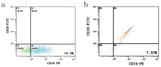
Recent studies have reported that cancer stem cells (CSCs) could be isolated from solid cancer cell lines, in which the purity of CSCs was higher than that from tumor tissues. Separation of CSCs from leukemic cell lines was rarely reported. In this study, CD34(+)CD38(-)stem-like cell subsets in human KG-1a leukemic cell line were enriched by cytotoxic agent 5-fluorouracil (5-FU). After 4 days incubation of KG-1a cell line with 5-FU (50 microg/ml), the CD34(+)CD38(-) subpopulation of cell lines was enriched more than 10 times. The enriched cells had proliferate potential in vitro, low level of RNA transcription and Hoechst 33342 dye efflux ability, accompanied by high expression of ATP-binding cassette transporter protein ABCG2. Our findings suggest that treatment with 5-FU offers an easy method to isolate leukemic stem-like subpopulation. It can facilitate studies of leukemic stem cell biology and the development of new therapeutic strategies.
Keywords: 5-fluorouracil; KG-1a; cell line; leukemia; stem cell.
Conflict of interest statement
Conflict of Interests: The authors have declared that no conflict of interest exists.
Figures


Similar articles
-
Determination of P-glycoprotein, MDR-related protein 1, breast cancer resistance protein, and lung-resistance protein expression in leukemic stem cells of acute myeloid leukemia.Cytometry B Clin Cytom. 2008 May;74(3):163-8. doi: 10.1002/cyto.b.20403. Cytometry B Clin Cytom. 2008. PMID: 18200595
-
Breast cancer resistance protein in drug resistance of primitive CD34+38- cells in acute myeloid leukemia.Clin Cancer Res. 2005 Mar 15;11(6):2436-44. doi: 10.1158/1078-0432.CCR-04-0212. Clin Cancer Res. 2005. PMID: 15788695
-
Relationships between multidrug resistance (MDR) and stem cell markers in human chronic myeloid leukemia cell lines.Leuk Res. 2010 Jun;34(6):757-62. doi: 10.1016/j.leukres.2009.11.004. Epub 2009 Dec 6. Leuk Res. 2010. PMID: 19969351
-
Side population cells in human cancers.Cancer Lett. 2008 Sep 8;268(1):1-9. doi: 10.1016/j.canlet.2008.03.048. Epub 2008 May 19. Cancer Lett. 2008. PMID: 18487012 Review.
-
Strategies for isolating and enriching cancer stem cells: well begun is half done.Stem Cells Dev. 2013 Aug 15;22(16):2221-39. doi: 10.1089/scd.2012.0613. Epub 2013 May 9. Stem Cells Dev. 2013. PMID: 23540661 Free PMC article. Review.
Cited by
-
CD24 Expression Is Increased in 5-Fluorouracil-Treated Esophageal Adenocarcinoma Cells.Front Pharmacol. 2017 May 30;8:321. doi: 10.3389/fphar.2017.00321. eCollection 2017. Front Pharmacol. 2017. PMID: 28611669 Free PMC article.
-
Can CD34+CD38- lymphoblasts, as likely leukemia stem cells, be a prognostic factor in B-cell precursor acute lymphoblastic leukemia in children?Front Pediatr. 2023 Aug 22;11:1213009. doi: 10.3389/fped.2023.1213009. eCollection 2023. Front Pediatr. 2023. PMID: 37675394 Free PMC article. Review.
-
Osteopontin b and c isoforms: Molecular Candidates Associated with Leukemic Stem Cell Chemoresistance in Acute Myeloid Leukemia.Asian Pac J Cancer Prev. 2017 Jun 25;18(6):1707-1715. doi: 10.22034/APJCP.2017.18.6.1707. Asian Pac J Cancer Prev. 2017. PMID: 28670893 Free PMC article.
-
Reciprocal Interactions of Leukemic Cells with Bone Marrow Stromal Cells Promote Enrichment of Leukemic Stem Cell Compartments in Response to Curcumin and Daunorubicin.Asian Pac J Cancer Prev. 2017 Mar 1;18(3):831-840. doi: 10.22034/APJCP.2017.18.3.831. Asian Pac J Cancer Prev. 2017. PMID: 28441794 Free PMC article.
-
OPN b and c Isoforms Doubtless Veto Anti-angiogenesis Effects of Curcumin in Combination with Conventional AML Regiment.Asian Pac J Cancer Prev. 2017 Sep 27;18(9):2591-2599. doi: 10.22034/APJCP.2017.18.9.2591. Asian Pac J Cancer Prev. 2017. PMID: 28952709 Free PMC article.
References
-
- Reya T, Morrison SJ, Clarke MF. et al.Stem cells, cancer, and cancer stem cells. Nature. 2001;414:105–11. - PubMed
-
- Bonnet D, Dick JE. Human acute myeloid leukemia is organized as a hierarchy that originates from a primitive hematopoietic cell. Nat Med. 1997;3:730–7. - PubMed
-
- Chan WI, Huntly BJ. Leukemia stem cells in acute myeloid leukemia. Semin Oncol. 2008;35:326–35. - PubMed
Publication types
MeSH terms
Substances
LinkOut - more resources
Full Text Sources
Other Literature Sources
Medical
Research Materials
