

Increased invasiveness and aggressiveness in breast epithelia with cytoplasmic p63 expression
- PMID: 20714441
- PMCID: PMC2920576
- DOI: 10.7150/ijbs.6.428
Increased invasiveness and aggressiveness in breast epithelia with cytoplasmic p63 expression
Abstract
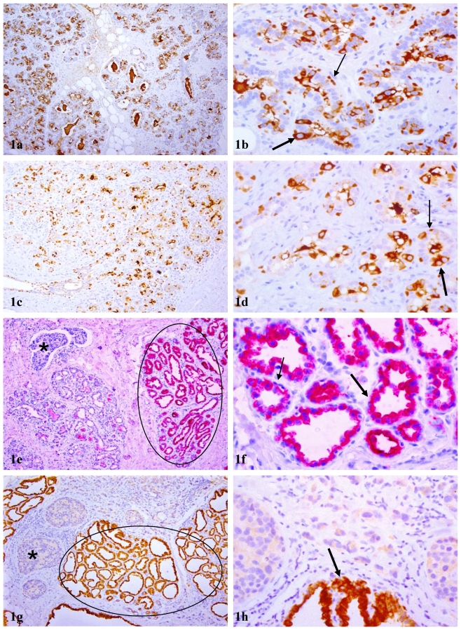
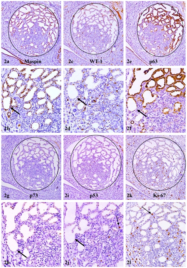
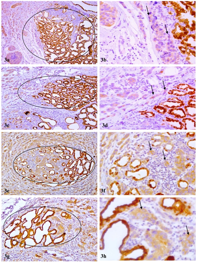
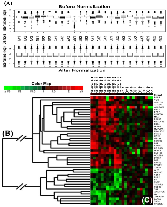
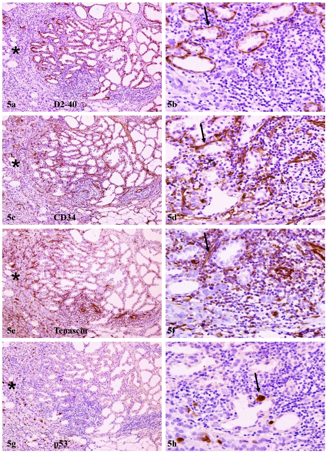
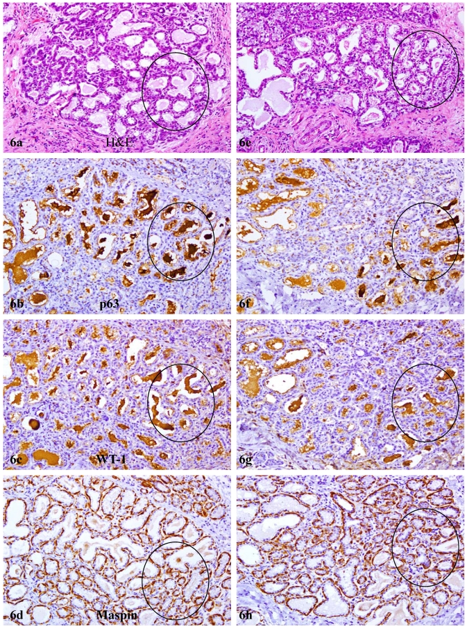
Our previous studies revealed that pregnancy associated breast cancer (PABC) had significantly reduced nuclear p63 expression in myoepithelia, while intense cytoplasmic p63 expression in associated epithelia. Our current study assessed these epithelia using immunohistochemistry with a panel of aggressiveness and invasiveness related markers and comparative genomic hybridization (array-CGH) with over 30,000 DNA probes. These epithelia showed several unique alterations, including (1) immunohistochemical and morphological resemblance to invasive cancer, (2) significant gain in copy numbers of DNA coding genes for morphogenesis, angiogenesis, and metastasis, and (3) significant loss in copy numbers of DNA coding genes for tumor suppressors, cell adhesion, and macromolecular complex assembly or intra-cellular trafficking. Detected array-CGH alterations correlated well with in vivo expression of a number of corresponding proteins tested. These findings suggest that aberrant sub-cellular localization of p63 expression in normal or hyperplastic appearing epithelial cells may significant contribute to increased invasiveness and aggressiveness of these cells.
Keywords: breast epithelia; invasiveness and aggressiveness.; p63 expression; pregnancy associated breast cancer.
Conflict of interest statement
Conflict of Interests: The authors have declared that no conflict of interest exists.
Figures
Similar articles
-
Aberrant p63 and WT-1 expression in myoepithelial cells of pregnancy-associated breast cancer: implications for tumor aggressiveness and invasiveness.Int J Biol Sci. 2009;5(1):82-96. doi: 10.7150/ijbs.5.82. Epub 2009 Jan 9. Int J Biol Sci. 2009. PMID: 19173015 Free PMC article.
-
Genomic signatures of pregnancy-associated breast cancer epithelia and stroma and their regulation by estrogens and progesterone.Horm Cancer. 2013 Jun;4(3):140-53. doi: 10.1007/s12672-013-0136-z. Epub 2013 Mar 12. Horm Cancer. 2013. PMID: 23479404 Free PMC article.
-
Hidden malignant cells within leukocyte aggregates: seeds for invasive and metastatic cancer?Cancer Epidemiol. 2011 Oct;35(5):475-9. doi: 10.1016/j.canep.2010.11.010. Epub 2011 Feb 2. Cancer Epidemiol. 2011. PMID: 21292584
-
p63 transcriptional regulation of epithelial integrity and cancer.Cell Cycle. 2007 Feb 1;6(3):240-5. doi: 10.4161/cc.6.3.3803. Epub 2007 Feb 3. Cell Cycle. 2007. PMID: 17297296 Review.
-
The guardians of the genome (p53, TA-p73, and TA-p63) are regulators of tumor suppressor miRNAs network.Cancer Metastasis Rev. 2010 Dec;29(4):613-39. doi: 10.1007/s10555-010-9257-9. Cancer Metastasis Rev. 2010. PMID: 20922462 Review.
Cited by
-
Rat mitochondrion-neuron focused microarray (rMNChip) and bioinformatics tools for rapid identification of differential pathways in brain tissues.Int J Biol Sci. 2011 Mar 29;7(3):308-22. doi: 10.7150/ijbs.7.308. Int J Biol Sci. 2011. PMID: 21494430 Free PMC article.
-
Structural Maintenance of Chromosomes protein 1: Role in Genome Stability and Tumorigenesis.Int J Biol Sci. 2017 Sep 3;13(8):1092-1099. doi: 10.7150/ijbs.21206. eCollection 2017. Int J Biol Sci. 2017. PMID: 28924389 Free PMC article. Review.
-
Pregnancy-associated breast cancer: the risky status quo and new concepts of predictive medicine.EPMA J. 2018 Feb 8;9(1):1-13. doi: 10.1007/s13167-018-0129-7. eCollection 2018 Mar. EPMA J. 2018. PMID: 29515683 Free PMC article. Review.
-
Protein deep sequencing applied to biobank samples from patients with pancreatic cancer.J Cancer Res Clin Oncol. 2015 Feb;141(2):369-80. doi: 10.1007/s00432-014-1817-x. Epub 2014 Sep 13. J Cancer Res Clin Oncol. 2015. PMID: 25216700 Free PMC article.
-
Biomarkers in an animal model for revealing neural, hematologic, and behavioral correlates of PTSD.J Vis Exp. 2012 Oct 10;(68):3361. doi: 10.3791/3361. J Vis Exp. 2012. PMID: 23093202 Free PMC article.
References
-
- Mathelin C, Annane K, Treisser A. et al.Pregnancy and post-partum breast cancer: a prospective study. Anticancer Res. 2008;28(4C):2447–2452. - PubMed
-
- Rodriguez AO, Chew H, Cress R. et al.Evidence of poorer survival in pregnancy-associated breast cancer. Obstet Gynecol. 2008;112(1):71–78. - PubMed
-
- Polyak K. Pregnancy and breast cancer: the other side of the coin. Cancer Cell. 2006;9(3):151–153. - PubMed
-
- Schedin P. Pregnancy-associated breast cancer and metastasis. Nat Rev Cancer. 2006;6(4):281–191. - PubMed
-
- Sternlight MD, Barsky SH. The myoepithelial defense: a host defense against cancer. Med Hypotheses. 1997;48:37– 46. - PubMed
Publication types
MeSH terms
Substances
Grants and funding
LinkOut - more resources
Full Text Sources
Medical
