Review
. 2010 Nov;120(5):567-84.
doi: 10.1007/s00401-010-0736-4. Epub 2010 Aug 17.

Molecular diagnostics of gliomas: state of the art

Affiliations
Review

Molecular diagnostics of gliomas: state of the art

Markus J Riemenschneider et al. Acta Neuropathol. 2010 Nov.

Abstract

Modern neuropathology serves a key function in the multidisciplinary management of brain tumor patients. Owing to the recent advancements in molecular neurooncology, the neuropathological assessment of brain tumors is no longer restricted to provide information on a tumor's histological type and malignancy grade, but may be complemented by a growing number of molecular tests for clinically relevant tissue-based biomarkers. This article provides an overview and critical appraisal of the types of genetic and epigenetic aberrations that have gained significance in the molecular diagnostics of gliomas, namely deletions of chromosome arms 1p and 19q, promoter hypermethylation of the O6-methylguanine-methyl-transferase (MGMT) gene, and the mutation status of the IDH1 and IDH2 genes. In addition, the frequent oncogenic aberration of BRAF in pilocytic astrocytomas may serve as a novel diagnostic marker and therapeutic target. Finally, this review will summarize recent mechanistic insights into the molecular alterations underlying treatment resistance in malignant gliomas and outline the potential of genome-wide profiling approaches for increasing our repertoire of clinically useful glioma markers.


Figures

Fig. 1
Fig. 1
Summary of most frequent molecular alterations in astrocytic, oligodendroglial, and oligoastrocytic gliomas. Primary glioblastomas, the most common gliomas in adults, show complex chromosomal, genetic, and epigenetic alterations targeting genes involved in important cellular pathways, namely the receptor tyrosin kinase/mitogen-activated protein kinase/phosphoinositol 3-kinase pathway (e.g. EGFR, MET, PDGFRA, ERBB2, NF1, PTEN, PIK3R1, PI3KCA, CTMP), the p53 pathway (e.g. TP53, p14 ARF , MDM2, MDM4), and the pRb1 pathway (e.g. CDKN2A, CDKN2B, CDK4, CDK6, RB1). In addition, primary glioblastomas frequently show monosomy 10, trisomy 7 and gains of 19q and 20q. Diffuse WHO grade II and III astrocytic, oligodendroglial and oligoastrocytic gliomas and secondary glioblastomas frequently carry mutations in IDH1 or IDH2, suggesting that they share a common, yet to be defined cell of origin. Diffuse astrocytic gliomas often carry additional TP53 mutations, while oligodendroglial tumors are characterized by 1p/19q deletion. Most oligoastrocytomas have either of these alterations. Molecular changes associated with progression to anaplastic glioma include 9p losses and inactivation of the CDKN2A, p14 ARF and CDKN2B genes on 9p21 as well as other changes, while progression to secondary glioblastoma is associated with frequent loss of 10q and DCC loss of expression among others. The majority of pilocytic astrocytomas are characterized by duplication/fusion or point mutation of the BRAF gene on 7q34, while other genomic aberrations are rare
Fig. 2
Fig. 2
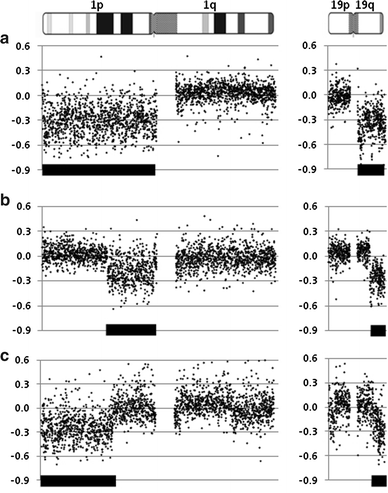
Different types of 1p and 19q losses as detected by array-CGH. Genomic profiles were obtained using tiling-resolution arrays containing 32,447 human BAC clones. Profiles are shown for chromosomes 1 and 19. On the X axis, BACs are aligned according to their physical mapping positions from the 1p telomere to the 1q telomere (n = 2,365) and from the 19p telomere to the 19q telomere (n = 735). On the the Y axis, the log2-transformed and normalized test:reference intensity ratios [“2Log(T/R)”]) are represented. Centromeric and heterochromatic regions are not evaluated using array-CGH resulting in absence of ratios (i.e. spots) in these regions and thereby visually separating the p-arm from the q-arm. Losses detected are indicated by a bar shown on the bottom of the ratio profiles, representing a complete chromosomes 1p and 19q loss (a), a loss of 1p11-p31.1, and 19q13.31-19qter (b), and a loss of 1pter-1p31.2 and 19q13.32-19qter (c)
Fig. 3
Fig. 3
Distribution of the four clinically relevant molecular alterations according to glioma entity. Colored squares indicate that the particular aberration is frequent in the respective tumor entity, i.e., usually detectable in 40% or more of the cases. Uncolored squares indicate that the aberration is rare in the respective tumor entity, i.e., usually restricted to <10% of the cases (except for MGMT promoter methylation being reported in approximately 20% of the pilocytic astrocytomas). Note that oligodendrogliomas and anaplastic oligodendrogliomas are characterized by the frequent coincidence of 1p/19q deletions, IDH1 or IDH2 mutation and MGMT promoter methylation (red squares). The same applies for oligoastrocytic tumors, although the frequency of 1p/19q deletion is less common when compared with “pure” oligodendrogliomas, also depending on the stringency of the histological classification used for mixed gliomas. IDH1 mutation and MGMT promoter methylation is frequent in diffuse astrocytomas, anaplastic astrocytomas and secondary glioblastomas (green squares). In contrast, primary glioblastomas rarely carry 1p/19q deletions and IDH1 mutations, while MGMT promoter methylation is found in approximately 40% of the cases (blue square). Pilocytic astrocytomas are uniquely characterized by BRAF alterations in more than 60% of the cases (orange square)

Similar articles

Cited by

References

    1. Antonelli M, Buttarelli FR, Arcella A, Nobusawa S, Donofrio V, Oghaki H and Giangaspero F (2010) Prognostic significance of histological grading, p53 status, YKL-40 expression, and IDH1 mutations in pediatric high-grade gliomas. J Neurooncol [Epub ahead of print] - PubMed
    1. Balss J, Meyer J, Mueller W, Korshunov A, Hartmann C, von Deimling A. Analysis of the IDH1 codon 132 mutation in brain tumors. Acta Neuropathol. 2008;116:597–602. - PubMed
    1. Bar EE, Lin A, Tihan T, Burger PC, Eberhart CG. Frequent gains at chromosome 7q34 involving BRAF in pilocytic astrocytoma. J Neuropathol Exp Neurol. 2008;67:878–887. - PubMed
    1. Barker FG, 2nd, Simmons ML, Chang SM, Prados MD, Larson DA, Sneed PK, Wara WM, Berger MS, Chen P, Israel MA, Aldape KD. EGFR overexpression and radiation response in glioblastoma multiforme. Int J Radiat Oncol Biol Phys. 2001;51:410–418. - PubMed
    1. Bayley JP, Devilee P. Warburg tumours and the mechanisms of mitochondrial tumour suppressor genes. Barking up the right tree? Curr Opin Genet Dev. 2010;20:324–329. - PubMed