Real-time measurement of solute transport within the lacunar-canalicular system of mechanically loaded bone: direct evidence for load-induced fluid flow
- PMID: 20715178
- PMCID: PMC3179346
- DOI: 10.1002/jbmr.211
Real-time measurement of solute transport within the lacunar-canalicular system of mechanically loaded bone: direct evidence for load-induced fluid flow
Abstract
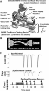
Since proposed by Piekarski and Munro in 1977, load-induced fluid flow through the bone lacunar-canalicular system (LCS) has been accepted as critical for bone metabolism, mechanotransduction, and adaptation. However, direct unequivocal observation and quantification of load-induced fluid and solute convection through the LCS have been lacking due to technical difficulties. Using a novel experimental approach based on fluorescence recovery after photobleaching (FRAP) and synchronized mechanical loading and imaging, we successfully quantified the diffusive and convective transport of a small fluorescent tracer (sodium fluorescein, 376 Da) in the bone LCS of adult male C57BL/6J mice. We demonstrated that cyclic end-compression of the mouse tibia with a moderate loading magnitude (-3 N peak load or 400 µε surface strain at 0.5 Hz) and a 4-second rest/imaging window inserted between adjacent load cycles significantly enhanced (+31%) the transport of sodium fluorescein through the LCS compared with diffusion alone. Using an anatomically based three-compartment transport model, the peak canalicular fluid velocity in the loaded bone was predicted (60 µm/s), and the resulting peak shear stress at the osteocyte process membrane was estimated (∼5 Pa). This study convincingly demonstrated the presence of load-induced convection in mechanically loaded bone. The combined experimental and mathematical approach presented herein represents an important advance in quantifying the microfluidic environment experienced by osteocytes in situ and provides a foundation for further studying the mechanisms by which mechanical stimulation modulates osteocytic cellular responses, which will inform basic bone biology, clinical understanding of osteoporosis and bone loss, and the rational engineering of their treatments.
Copyright © 2011 American Society for Bone and Mineral Research.
Figures


Similar articles
-
Quantifying load-induced solute transport and solute-matrix interaction within the osteocyte lacunar-canalicular system.J Bone Miner Res. 2013 May;28(5):1075-86. doi: 10.1002/jbmr.1804. J Bone Miner Res. 2013. PMID: 23109140 Free PMC article.
-
Does blood pressure enhance solute transport in the bone lacunar-canalicular system?Bone. 2010 Aug;47(2):353-9. doi: 10.1016/j.bone.2010.05.005. Epub 2010 May 13. Bone. 2010. PMID: 20471508 Free PMC article.
-
In situ measurement of solute transport in the bone lacunar-canalicular system.Proc Natl Acad Sci U S A. 2005 Aug 16;102(33):11911-6. doi: 10.1073/pnas.0505193102. Epub 2005 Aug 8. Proc Natl Acad Sci U S A. 2005. PMID: 16087872 Free PMC article.
-
Solute Transport in the Bone Lacunar-Canalicular System (LCS).Curr Osteoporos Rep. 2018 Feb;16(1):32-41. doi: 10.1007/s11914-018-0414-3. Curr Osteoporos Rep. 2018. PMID: 29349685 Free PMC article. Review.
-
Multiscale finite element modeling of mechanical strains and fluid flow in osteocyte lacunocanalicular system.Bone. 2020 Aug;137:115328. doi: 10.1016/j.bone.2020.115328. Epub 2020 Mar 20. Bone. 2020. PMID: 32201360 Free PMC article. Review.
Cited by
-
In vitro and in vivo approaches to study osteocyte biology.Bone. 2013 Jun;54(2):296-306. doi: 10.1016/j.bone.2012.09.040. Epub 2012 Oct 13. Bone. 2013. PMID: 23072918 Free PMC article. Review.
-
Regional differences in oxidative metabolism and mitochondrial activity among cortical bone osteocytes.Bone. 2016 Sep;90:15-22. doi: 10.1016/j.bone.2016.05.011. Epub 2016 May 31. Bone. 2016. PMID: 27260646 Free PMC article.
-
Mechanical Loading Differentially Affects Osteocytes in Fibulae from Lactating Mice Compared to Osteocytes in Virgin Mice: Possible Role for Lacuna Size.Calcif Tissue Int. 2018 Dec;103(6):675-685. doi: 10.1007/s00223-018-0463-8. Epub 2018 Aug 14. Calcif Tissue Int. 2018. PMID: 30109376 Free PMC article.
-
Spatial variations in the osteocyte lacuno-canalicular network density and analysis of the connectomic parameters.PLoS One. 2024 May 14;19(5):e0303515. doi: 10.1371/journal.pone.0303515. eCollection 2024. PLoS One. 2024. PMID: 38743675 Free PMC article.
-
Informing phenomenological structural bone remodelling with a mechanistic poroelastic model.Biomech Model Mechanobiol. 2016 Feb;15(1):69-82. doi: 10.1007/s10237-015-0735-4. Epub 2015 Nov 3. Biomech Model Mechanobiol. 2016. PMID: 26534771 Free PMC article.
References
-
- Bonewald LF. Osteocytes as dynamic multifunctional cells. Ann NY Acad Sci. 2007;1116:281–290. - PubMed
-
- Kogianni G, Noble BS. The biology of osteocytes. Curr Osteoporos Rep. 2007;5:81–86. - PubMed
-
- Piekarski K, Munro M. Transport mechanism operating between blood supply and osteocytes in long bones. Nature. 1977;269:80–82. - PubMed
-
- Hancox NM. Biology of Bone: Biological Structure and Function. Cambridge, United Kingdon: Cambridge University Press; 1972. p. viii.
-
- You LD, Weinbaum S, Cowin SC, Schaffler MB. Ultrastructure of the osteocyte process and its pericellular matrix. Anat Rec A Discov Mol Cell Evol Biol. 2004;278:505–513. - PubMed
Publication types
MeSH terms
Substances
Grants and funding
LinkOut - more resources
Full Text Sources
Miscellaneous

