. 2010 Oct;28(10):1893-904.
doi: 10.1002/stem.499.

Efficient generation of functional dopaminergic neurons from human induced pluripotent stem cells under defined conditions

Affiliations
Free PMC article

Efficient generation of functional dopaminergic neurons from human induced pluripotent stem cells under defined conditions

Andrzej Swistowski et al. Stem Cells. 2010 Oct.
Free PMC article

Abstract

Human induced pluripotent stem cells (iPSCs) reprogrammed from somatic cells represent a promising unlimited cell source for generating patient-specific cells for biomedical research and personalized medicine. As a first step, critical to clinical applications, we attempted to develop defined culture conditions to expand and differentiate human iPSCs into functional progeny such as dopaminergic neurons for treating or modeling Parkinson's disease (PD). We used a completely defined (xeno-free) system that we previously developed for efficient generation of authentic dopaminergic neurons from human embryonic stem cells (hESCs), and applied it to iPSCs. First, we adapted two human iPSC lines derived from different somatic cell types for the defined expansion medium and showed that the iPSCs grew similarly as hESCs in the same medium regarding pluripotency and genomic stability. Second, by using these two independent adapted iPSC lines, we showed that the process of differentiation into committed neural stem cells (NSCs) and subsequently into dopaminergic neurons was also similar to hESCs. Importantly, iPSC-derived dopaminergic neurons were functional as they survived and improved behavioral deficits in 6-hydroxydopamine-leasioned rats after transplantation. In addition, iPSC-derived NSCs and neurons could be efficiently transduced by a baculoviral vector delivering episomal DNA for future gene function study and disease modeling using iPSCs. We also performed genome-wide microarray comparisons between iPSCs and hESCs, and we derived NSC and dopaminergic neurons. Our data revealed overall similarity and visible differences at a molecular level. Efficient generation of functional dopaminergic neurons under defined conditions will facilitate research and applications using PD patient-specific iPSCs.


Figures

Figure 1
Figure 1
Generation of neural stem cells (NSCs) from induced pluripotent stem cell (iPSC) lines adapted to defined medium. iPSC line MR31 at passage 15 was adapted to a chemically defined medium StemPro. (A–D): Morphology (A) and expression of the pluripotent markers Tra 1-60 (B), Oct4 (C), and SSEA4 (D) in iPSCs that were cultured in StemPro for 10 passages. (E–H): Generation of NSCs in defined conditions. Neural tube-like rosette structures (E) were formed in the center of the iPSC colonies after 12 days of differentiation. A monolayer of homogeneous NSCs (F) coexpressed Sox1 and nestin (G), and Musashi (H). (I–L): iPSC-deriver NSCs retained the capacity to differentiate into neurons (I–J), astrocytes (K), and oligodendrocytes (L). Abbreviation: GFAP, Glial fibrillary acidic protein.
Figure 2
Figure 2
Efficient differentiation of induced pluripotent stem cell (iPSC)-derived NSCs into dopaminergic neurons. (A–D): iPSC-derived NSCs differentiated into midbrain dopaminergic neurons in defined media as seen by immunocytochemistry. The majority of the cells expressed β-III-tubulin and TH after 5 weeks of differentiation (A–C). Coexpression of A9 marker Girk2 in TH+ dopaminergic neurons (D). Differential expression of dopaminergic markers in dopaminergic neurons compared with NSCs by quantitative polymerase chain reaction (E). All the examined markers were upregulated in dopaminergic populations compared with NSCs. Abbreviations: AADC, Aromatic L-Amino Acid Decarboxylase; DAT, dopamine transporter; NSC, neural stem cell; TH, tyrosine hydroxylase.
Figure 3
Figure 3
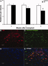
Transplantation of induced pluripotent stem cell (iPSC)-derived cells into 6-hydroxydopamine (6-OHDA) Parkinson's disease (PD) rats. iPSC-derived dopaminergic neurons engrafted and ameliorated behavioral deficits in a PD model. (A): Amphetamine-induced rotations in 6-OHDA rats grafted with iPSC-derived neurons (20 days after the neural stem cell stage) showed significant rotational improvement 12 weeks after transplantation. (B–E): Histological analysis of a representative brain section showed that donor cells (human antigen-immunopositive cells) coexpressing TH survived in the graft sites 12 weeks post-transplantation. Abbreviation: TH, tyrosine hydroxylase.
Figure 4
Figure 4
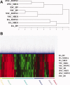
Whole genomic analysis of iPSCs at different stages of dopaminergic differentiation by Illumina bead microarray. A total of nine samples (undifferentiated human embryonic stem cell [hESC] line H9, iPSC lines MR31 and MMW2, NSCs derived from H9, MR31, and MMW2, and dopaminergic neurons derived from H9, MR31, and MMW2) were analyzed by Illumina array and the results demonstrated similarities between hESC and iPSCs. (A): Dendrogram of unsupervised one-way hierarchical clustering analysis of global gene expression data in hESCs/iPSCs, NSCs, and dopaminergic neurons derived from hESCs/iPSCs. (B): Unsupervised two-way hierarchical cluster analysis of differentially expressed genes illustrated in a heat map. The samples include three groups, hESCs/iPSCs, NSCs, and dopaminergic neurons. Expression values are presented as the log2 signal value of the given gene. Abbreviations: DA, dopaminergic; iPSC, induced pluripotent stem cell; NSC, neural stem cell.
Figure 5
Figure 5
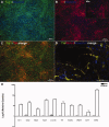
Efficient transduction of induced pluripotent stem cell (iPSC)-derived neural stem cells (NSCs) and neurons by baculoviral vector. iPSC-derived NSCs and neurons were transduced by a baculoviral vector carrying a GFP driven by the CMV promoter. (A–L): Fluorescence microscopy showed strong expression of GFP in iPSC-derived NSCs (87% of total cell were GFP+ by FACS) 24 hours post-transduction or day 36 neurons (90.3% of total cell were GFP+ by FACS) 2 days after transduction. Expression of GFP in human embryonic stem cell (hESC)-derived neurons was shown for comparison (E, F). Immunostaining of β-III tubulin confirmed that the majority of neurons coexpressed GFP in both iPSC-derived neurons (G–I) and hESC-derived neurons (J–L). Abbreviation: GFP, Green Fluorescent Protein; CMV, Cytomegalovirus; FACS, Fluorescence-Activated Cell Sorting.

Similar articles

Cited by

References

    1. Yan Y, Yang D, Zarnowska ED, et al. Directed differentiation of dopaminergic neuronal subtypes from human embryonic stem cells. Stem Cells. 2005;23:781–790. - PMC - PubMed
    1. Zeng X, Cai J, Chen J, et al. Dopaminergic differentiation of human embryonic stem cells. Stem Cells. 2004;22:925–940. - PubMed
    1. Perrier AL, Tabar V, Barberi T, et al. Derivation of midbrain dopamine neurons from human embryonic stem cells. Proc Natl Acad Sci USA. 2004;101:12543–12548. - PMC - PubMed
    1. Thomson JA, Itskovitz-Eldor J, Shapiro SS, et al. Embryonic stem cell lines derived from human blastocysts. Science. 1998;282:1145–1147. - PubMed
    1. Swistowski A, Peng J, Han Y, et al. Xeno-free defined conditions for culture of human embryonic stem cells, neural stem cells and dopaminergic neurons derived from them. Plos One. 2009;4:e6233. - PMC - PubMed

Publication types