

doi: 10.1002/anie.201002906.
Backbone dynamics of cyclotide MCoTI-I free and complexed with trypsin
Affiliations
- PMID: 20715250
- PMCID: PMC2944905
- DOI: 10.1002/anie.201002906
Item in Clipboard
Backbone dynamics of cyclotide MCoTI-I free and complexed with trypsin
Angew Chem Int Ed Engl.
.
Erratum in
- Angew Chem Int Ed Engl. 2011 Jul 25;50(31):6948-9
No abstract available
Figures
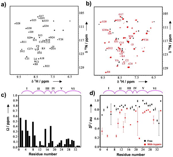
NMR analysis of the backbone dynamic of free and trypsin bound MCoTI-I. (a) NMR {15N,1H}-HSQC spectrum of free MCoTI-I. Chemical shift assignments of the backbone amides are indicated. (b) Overlay of the {15N,1H}-HSQC spectra of free (black) and trypsin bound MCoTI-I (red). Residues with large average amide chemical shift differences between two different states ( > 0.3 ppm) are indicated. Peaks that are broadened in trypsin bound MCoTI-I are indicated by grey circles. (c) Average amide chemical shift difference for all the assigned residues in free and trypsin bound MCoTI-I. Chemical shift difference was calculated as: ΔΩ = [(ΔΩNH2+0.04ΔΩN2)/2]½ , where ΔΩNH and ΔΩN are the changes in the amide proton and nitrogen chemical shifts (ppm), respectively. (d) The order parameter, S2, for the free (black) and the trypsin bound MCoTI-I (red). S2 value is a measure of backbone flexibility and represents the degree of angular restriction of N-H vector in the molecular frame. The MCoTI-I loops are shown on top of panels C and D. Small unassigned peaks in both free and trypsin bound spectra of MCoTI-I are from a minor conformation of the protein due to a known isomerization of the backbone at an Asp-Gly sequence in loop 6 of MCoTI-I.
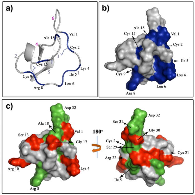
Trypsin binding to MCoTI-I affects the MCoTI-I backbone dynamics. Ribbon (a) and surface (b) diagrams of the trypsin-MCoTI-I interaction map. Red arabic numbers indicate the positions of the MCoTI-I loops. The MCoTI-I residues with a large chemical shift difference (>0.3 ppm) are in blue. (c) Changes in the MCoTI-I order parameter due to binding to trypsin. Residues with Sf2 - Sb2 > 0.2, where Sf/b2 is the order parameter of the free or trypsin bound MCoTI-I, respectively, are depicted in red. MCoTI-I residues that were broadened in {15N,1H}-HSQC due to binding to trypsin are shown in green. We used a structure of free MCoTI-II (PDB code: 1IB9)[6] to illustrate the changes of MCoTI-I dynamics due to trypsin binding.
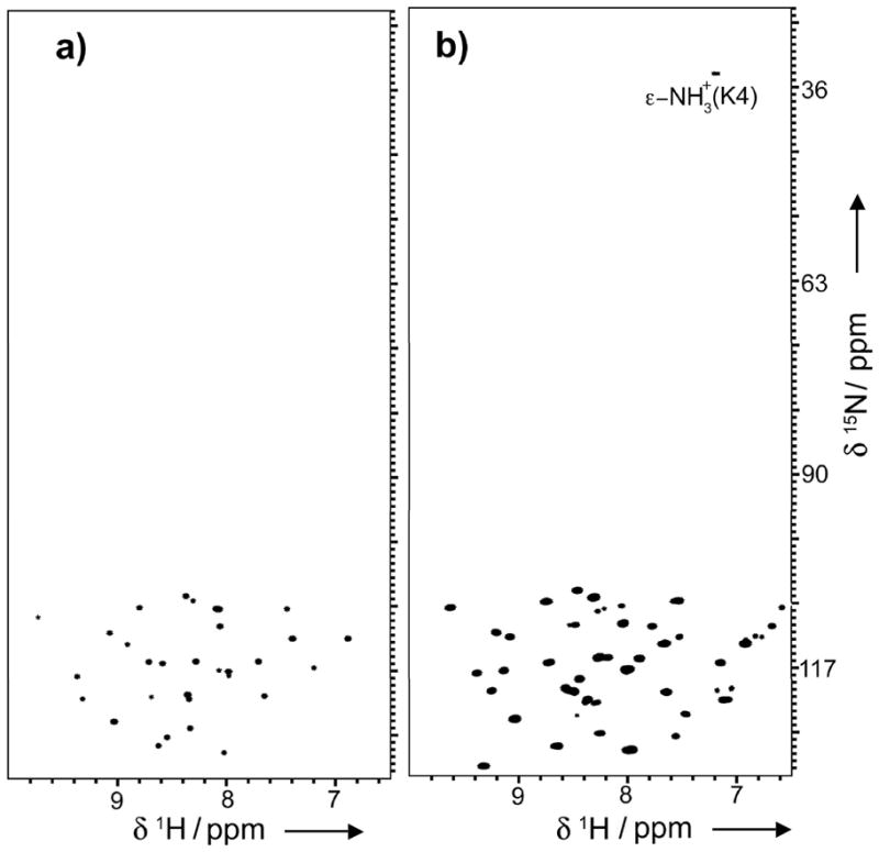
ε-NH3+ of Lys4 is protected from fast exchange with the solvent in trypsin bound MCoTI-I. {15N,1H}-HSQC spectra of free (a) and trypsin-bound MCoTI-I (b) were collected at RT with the 15N-carrier position at 82 ppm and 15N rf field strengths of 5.2 kHz for 90° and 180° pulses and 1.2 kHz for composite decoupling during acquisition.
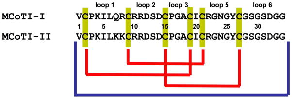
Primary structure and disulfide connectivities of MCoTI cyclotides. Blue and yellow connectors represent peptide and disulfide bonds, respectively.
Similar articles
-
Computational analysis of the MCoTI-II plant defence knottin reveals a novel intermediate conformation that facilitates trypsin binding.Sci Rep. 2016 Mar 15;6:23174. doi: 10.1038/srep23174. Sci Rep. 2016. PMID: 26975976 Free PMC article.
-
Cyclotides, a versatile ultrastable micro-protein scaffold for biotechnological applications.Bioorg Med Chem Lett. 2017 Dec 1;27(23):5089-5099. doi: 10.1016/j.bmcl.2017.10.051. Epub 2017 Oct 21. Bioorg Med Chem Lett. 2017. PMID: 29110985 Free PMC article. Review.
-
Structural insights into the role of the cyclic backbone in a squash trypsin inhibitor.J Biol Chem. 2013 Dec 13;288(50):36141-8. doi: 10.1074/jbc.M113.528240. Epub 2013 Oct 29. J Biol Chem. 2013. PMID: 24169696 Free PMC article.
-
Knots in rings. The circular knotted protein Momordica cochinchinensis trypsin inhibitor-II folds via a stable two-disulfide intermediate.J Biol Chem. 2006 Mar 24;281(12):8224-32. doi: 10.1074/jbc.M513399200. Epub 2006 Jan 23. J Biol Chem. 2006. PMID: 16547012
-
Oxidative folding of the cystine knot motif in cyclotide proteins.Protein Pept Lett. 2005 Feb;12(2):147-52. doi: 10.2174/0929866053005863. Protein Pept Lett. 2005. PMID: 15723640 Review.
Cited by
-
Chemical synthesis, backbone cyclization and oxidative folding of cystine-knot peptides: promising scaffolds for applications in drug design.Molecules. 2012 Oct 24;17(11):12533-52. doi: 10.3390/molecules171112533. Molecules. 2012. PMID: 23095896 Free PMC article. Review.
-
Expression of fluorescent cyclotides using protein trans-splicing for easy monitoring of cyclotide-protein interactions.Angew Chem Int Ed Engl. 2013 Mar 11;52(11):3126-31. doi: 10.1002/anie.201209219. Epub 2013 Jan 15. Angew Chem Int Ed Engl. 2013. PMID: 23322720 Free PMC article. No abstract available.
-
Intein applications: from protein purification and labeling to metabolic control methods.J Biol Chem. 2014 May 23;289(21):14512-9. doi: 10.1074/jbc.R114.552653. Epub 2014 Apr 2. J Biol Chem. 2014. PMID: 24700459 Free PMC article. Review.
-
Design of a MCoTI-Based Cyclotide with Angiotensin (1-7)-Like Activity.Molecules. 2016 Jan 26;21(2):152. doi: 10.3390/molecules21020152. Molecules. 2016. PMID: 26821010 Free PMC article.
-
The Potential of the Cyclotide Scaffold for Drug Development.Biomedicines. 2019 Apr 19;7(2):31. doi: 10.3390/biomedicines7020031. Biomedicines. 2019. PMID: 31010257 Free PMC article. Review.
References
-
- Daly NL, Rosengren KJ, Craik DJ. Adv Drug Deliv Rev. 2009;61:918. - PubMed
-
- Craik DJ, Simonsen S, Daly NL. Curr Opin Drug Discov Devel. 2002;5:251. - PubMed
-
- Craik DJ, Cemazar M, Wang CK, Daly NL. Biopolymers. 2006;84:250. - PubMed
-
- Hernandez JF, Gagnon J, Chiche L, Nguyen TM, Andrieu JP, Heitz A, Trinh Hong T, Pham TT, Le Nguyen D. Biochemistry. 2000;39:5722. - PubMed
-
- Heitz A, Hernandez JF, Gagnon J, Hong TT, Pham TT, Nguyen TM, Le-Nguyen D, Chiche L. Biochemistry. 2001;40:7973. - PubMed
Publication types
MeSH terms
Substances
Grants and funding
LinkOut - more resources
Full Text Sources
Other Literature Sources
