

doi: 10.1002/cphc.201000528.
Chemistries for patterning robust DNA microbarcodes enable multiplex assays of cytoplasm proteins from single cancer cells
Affiliations
- PMID: 20715281
- PMCID: PMC3681607
- DOI: 10.1002/cphc.201000528
Item in Clipboard
Chemistries for patterning robust DNA microbarcodes enable multiplex assays of cytoplasm proteins from single cancer cells
Chemphyschem.
.
No abstract available
Figures
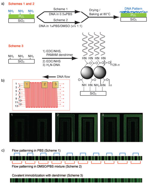
a) Surface treatment schemes. b) Design of the DNA patterning device (top) and fluorescence image of DNAs filled into the channel (still in solution). Outer five channels are filled with DNAs in 1:1 mixture of PBS and water (Scheme 1). The five inner are filled with DNA in a 1:1 mixture of PBS and DMSO (Scheme 2). Three channels in between are left empty for visualization. c) Fluorescence images of patterned DNAs by three schemes.
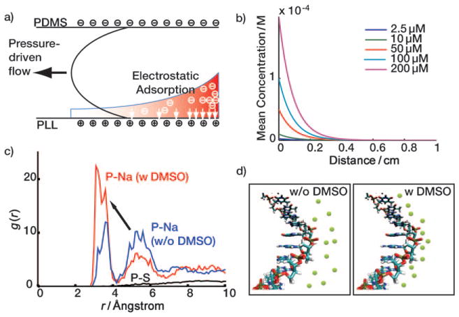
Electrostatic adsorption of DNAs on PLL surface and DMSO effect. a) The filling step. b) Simulation result of electrostatic adsorption of DNAs to PLL surface. c) Molecular simulation of the DMSO effect: the radial distribution function of P atom of the phosphate group and the sodium ions. The presence of DMSO pumps sodium ions from the 2nd shell to the 1st shell (arrow). d) Schematics for DMSO effect. Green circles represent sodium ions.
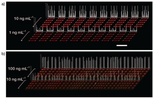
Contrast-enhanced raw data extracted from multi-protein calibration experiments performed on a substrate prepared according to a) Scheme 2 and b) Scheme 3. Each red bar represents a unique protein measurement, and is clustered with up to ten additional proteins (for Scheme 2). The clusters become symmetrical due to the winding nature of the barcode pattern, so that each cluster actually contains two measurements of each protein. Clustering is less evident in (b) because lower-density barcode pattern was employed. Recombinant proteins were analyzed across five discrete channels per concentration for (a) and four discrete channels per concentration for (b); quantitative data for statistical analysis was extracted from all the repeats in each of the channels. By utilizing identical DEAL cocktails followed by identical standard protein cocktails, the reproducibility was also checked. The identical signal patterns within individual channels and between channels of similar concentrations demonstrate the good uniformity and quality of DNA barcodes. Signal intensity profiles sampled from one analysis channel per concentration are quantified in white. Scale bar: 2 mm.
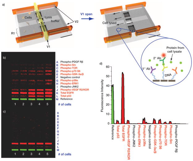
a) Schematic representation of the single-cell, intracellular protein analysis device. Single or few cells are incubated in an isolated chamber under varying stimuli. Intracellular proteins are assayed by introducing a pre-aliquoted lysis buffer, whereupon the released proteins bind to the DEAL (DNA-labeled antibody) barcode within the chamber. V1: valve for lysis buffer control, V2: valve for isolated chamber formation, and R1: DNA barcode array converted into DEAL antibody array. b), c) Contrast-enhanced images of developed barcode assays highlight the benefits of using Scheme 2 (b) versus Scheme 1 (c). Protein names listed in red font correspond to those which were detected using Scheme 2 barcodes. d) Representative fluorescence intensity profile from the single-cell lysate of (b).
References
Publication types
MeSH terms
Substances
Grants and funding
LinkOut - more resources
Full Text Sources
Other Literature Sources
