. 2011 Jan;15(1):31-7.
doi: 10.1111/j.1582-4934.2010.01152.x.

Membrane-directed molecular assembly of the neuronal SNARE complex

Affiliations

Membrane-directed molecular assembly of the neuronal SNARE complex

Won Jin Cho et al. J Cell Mol Med. 2011 Jan.

Abstract

Since the discovery and implication of N-ethylmaleimide-sensitive factor (NSF)-attachment protein receptor (SNARE) proteins in membrane fusion almost two decades ago, there have been significant efforts to understand their involvement at the molecular level. In the current study, we report for the first time the molecular interaction between full-length recombinant t-SNAREs and v-SNARE present in opposing liposomes, leading to the assembly of a t-/v-SNARE ring complex. Using high-resolution electron microscopy, the electron density maps and 3D topography of the membrane-directed SNARE ring complex was determined at nanometre resolution. Similar to the t-/v-SNARE ring complex formed when 50 nm v-SNARE liposomes meet a t-SNARE-reconstituted planer membrane, SNARE rings are also formed when 50 nm diameter isolated synaptic vesicles (SVs) meet a t-SNARE-reconstituted planer lipid membrane. Furthermore, the mathematical prediction of the SNARE ring complex size with reasonable accuracy, and the possible mechanism of membrane-directed t-/v-SNARE ring complex assembly, was determined from the study. Therefore in the present study, using both lipososome-reconstituted recombinant t-/v-SNARE proteins, and native v-SNARE present in isolated SV membrane, the membrane-directed molecular assembly of the neuronal SNARE complex was determined for the first time and its size mathematically predicted. These results provide a new molecular understanding of the universal machinery and mechanism of membrane fusion in cells, having fundamental implications in human health and disease.


Figures

Fig 1
Fig 1
Schematic flow diagram establishing membrane-associated SNARE complex assembly and its structural evaluation using AFM and EM. (A) Preteoliposomes of a specific size containing v-SNARE can be exposed to t-SNARE-reconstituted lipid membrane supported by a mica surface. The interaction results in the formation of a highly stable t-/v-SNARE ring complex which can be observed using AFM following removal of the vesicle. (B) Similarly, proteoliposomes of a certain size, one reconstituted with v-SNARE and the other with t-SNARE, can interact (C, D) to form membrane-directed t-/v-SNARE complex. However, in this situation, a single t-SNARE vesicle (red) is physically limited to establish contact with a maximum of 12 (blue) equal-size v-SNARE vesicles, and vice versa. This allows for approximately no more than 6% of the total surface area of one vesicle to interact with others, translating the membrane-directed t-/v-SNARE complex to be just around 6% of the total. In order to observe this membrane-directed SNARE complex, the liposomes are solubilized using detergent, and the stable t-/v-SNARE ring complex formed as a result of membrane-directed assembly, is then imaged using AFM and EM. (C) Schematic diagram depicting the possible molecular mechanism of SNARE ring complex formation, when t-SNARE-vesicles and V-SNARE-vesicles meet. The process may occur due to a progressive recruitment of t-/v-SNARE pairs as the opposing vesicles are pulled toward each other, until a complete ring is established, preventing any further recruitment of t-/v-SNARE pairs to the complex. The top panel is a side view of two vesicles (one t-SNARE-reconstituted, and the other v-SNARE reconstituted) interacting to form a single t-/v-SNARE complex, leading progressively (from left to right) to the formation of the ring complex. The lower panel is a top view of the two interacting vesicles.
Fig 2
Fig 2
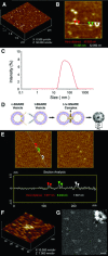
Atomic force micrographs of PC:PS vesicles extruded through a membrane of 50 nm pore size. (A) Low-resolution image of PC:PS vesicles in buffer on mica surface. (B) High-resolution image of PC:PS vesicles in buffer on mica surface. Section analysis through three PC:PS vesicles demonstrating each of them to measure approximately 50 nm in diameter. (C) PCS demonstrating the average vesicle size to be approximately 50 nm. (D) Schematic drawing of a v-SNARE proteoliposome, shown interacting with a t-SNARE proteoliposome, to form a t-/v-SNARE ring complex (RC) having a central pore (CP) or channel. (E) Low-resolution atomic force micrographs of the SNARE ring complex on mica surface in buffer. Note in the AFM section analysis of the 7.5–8.466 nm diameter t-/v-SNARE ring complexes formed when approximately 50 nm diameter t-SNARE-reconstituted vesicles and 50 nm diameter v-SNARE-reconstituted vesicles meet. (F) A high-resolution AFM image of the t-/v-SNARE ring complex. (G) Low-resolution electron micrograph of the t-/v-SNARE ring complexes (Bar = 20 nm). The inset is an EM micrograph of a 7.5 nm diameter t-/v-SNARE ring complex within a 10 nm box.
Fig 3
Fig 3
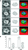
Electron micrograph (A, left column), electron density map (A, centre column), and 3D contour map (A, right column) of isolated t-/v-SNARE ring complex formed when 50 nm diameter t-SNARE-reconstituted vesicles and 50 nm diameter v-SNARE-reconstituted vesicles meet (Bar 5 7 nm). (B) Vertical bars indicate SEM. Note the 8.28 ± 0.85 nm diameter hydrated SNARE ring complex (RC) having a 2.84 6 0.57 nm diameter central pore (CP) within. In contrast, dehydrated SNARE ring complex from the same preparation, examined using EM, measure 7.31 ± 0.69 nm, and its central pore 2.02 ± 0.41 nm diameter.
Fig 4
Fig 4
Electron micrographs (A), electron density maps (B), and 3D contour maps (C) of isolated t-/v-SNARE ring complexes formed when approximately 50 nm diameter t-SNARE-reconstituted vesicles and 50 nm diameter v-SNARE-reconstituted vesicles interact. 3D topography of t/v-SNARE obtained from their corresponding electron maps. The colours from blue, through green to red; and from black, through grey to white, correspond to the protein image density from lowest to the highest. The highest peak in each image represents 30 pixels (1.4 A/pixel). Box size = 15 nm. (D–F) Wall-eye stereo view of the 3D topographies of SNARE ring complexes is presented. (G) Histogram of the intensity of a t-/v-SNARE ring complex is shown. The similarity between the histograms may correspond to the similarity between the topological structures of the complex. (H) One-dimensional intensity distribution along the horizontal direction of a SNARE ring complex, similar to an AFM section analysis. The intensity distribution represents an average of 9 pixels wide stripe taken across the centre of a SNARE ring complex. Note the central channel in the intensity distribution map.
Fig 5
Fig 5
The possible establishment of a leak-proof SNARE ring complex channel is demonstrated (A). Size of the t-/v-SNARE ring complex is directly proportional to the size of the SNARE-associated vesicle (B). Different sizes of v-SNARE-associated vesicles, when interacting with t-SNARE-associated membrane (○), demonstrate the SNARE ring size to be directly proportional to the vesicle size. When a 50 nm diameter v-SNARE-reconstituted vesicle interacts with a t-SNARE-reconstituted membrane, a 11 nm diameter t-/v-SNARE ring complex is formed. Similarly, the present study demonstrates that when a 50 nm diameter v-SNARE-reconstituted vesicle, interacts with a 50 nm diameter t-SNARE-reconstituted vesicle, an 8 nm diameter t-/v-SNARE ring complex is established (♦). Analogous to the 11 nm diameter t-/v-SNARE ring complexes formed when 50 nm v-SNARE vesicles meet a t-SNARE-reconstituted planer membrane (B), approximately 11 nm diameter t-/v-SNARE ring complexes are formed when 50 nm diameter SVs meet a t-SNARE-reconstituted planer membrane (C, D).

References

    1. Oyler GA, Higgins GA, Hart RA, et al. The identification of a novel synaptosomal-associated protein, SNAP-25, differentially expressed by neuronal subpopulations. J Cell Biol. 1989;109:3039–52. - PMC - PubMed
    1. Bennett MK, Calakos N, Scheller RH. Syntaxin: a synaptic protein implicated in docking of synaptic vesicles at presynaptic active zones. Science. 1992;257:255–9. - PubMed
    1. Trimble WS, Cowam DM, Scheller RH. VAMP-1: a synaptic vesicle-associated integral membrane protein. Proc Natl Acad Sci USA. 1988;85:4538–42. - PMC - PubMed
    1. Cho SJ, Kelly M, Rognlien KT, et al. SNAREs in opposing bilayers interact in a circular array to form conducting pores. Biophys J. 2002;83:2522–7. - PMC - PubMed
    1. Cho WJ, Jeremic A, Jena BP. Size of supramolecular SNARE complex: membrane-directed self-assembly. J Am Chem Soc. 2005;127:10156–7. - PMC - PubMed

Publication types