

Changes of anabolic processes at the cellular and molecular level in chronic wounds under topical negative pressure can be revealed by transcriptome analysis
- PMID: 20716124
- PMCID: PMC3823200
- DOI: 10.1111/j.1582-4934.2010.01147.x
Changes of anabolic processes at the cellular and molecular level in chronic wounds under topical negative pressure can be revealed by transcriptome analysis
Abstract
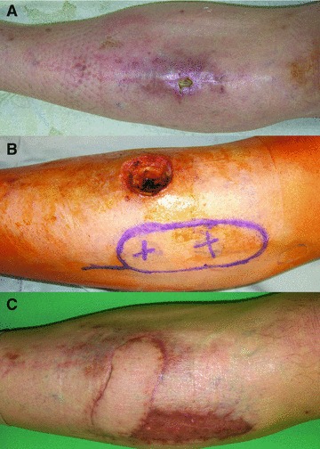
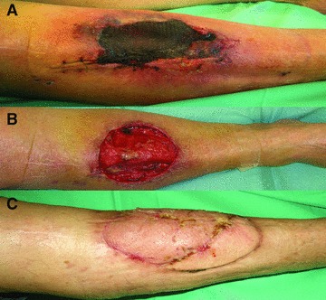
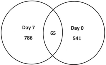
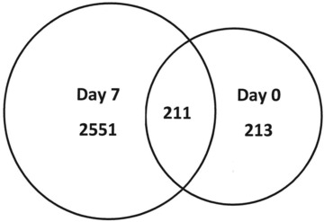
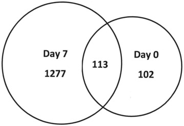
Chronic wounds--as defined by the World Union of Wound Healing Societies (WUWHS)--are a considerable worldwide health care expense and impair quality of life. In order for chronic wounds to heal, these wounds must be transformed to a more acute state to begin the healing process. Topical negative pressure (TNP) with reticulated open cell foam (ROCF) is known to promote healing in certain types of chronic wounds. However, little is known about changes at the cellular or molecular level in wounds under various treatments, especially under the physical forces induced to tissue by TNP. In the current study, chronic wound samples were obtained during routine wound debridements prior to treatment and 7-12 days after initiating TNP with a continuous setting at -125 mmHg. Whole genome transcriptome microarray analyses were performed on samples to better understand how TNP with ROCF affects these types of wounds. It was found that more genes were expressed following TNP with ROCF as compared to before therapy and to normal, non-wounded tissue. In this study, we show that TNP with ROCF transforms the chronic wound from its inflammation (non-healing) state into more of a progressive, healing phenotype from a molecular point of view with expression of genes that are commonly associated with these terms.
© 2011 The Authors Journal of Cellular and Molecular Medicine © 2011 Foundation for Cellular and Molecular Medicine/Blackwell Publishing Ltd.
Figures
References
-
- Argenta LC, Morykwas MJ. Vacuum-assisted closure: a new method for wound control and treatment: clinical experience. Ann Plast Surg. 1997;38:563–76. - PubMed
-
- Joseph E, Hamori CA, Bergman S, et al. A prospective, randomized trial of vacuum-assisted closure versus standard therapy of chronic nonhealing wounds. Wounds. 2000;12:60–7.
-
- Short B, Claxton M, Armstrong DG. How to use VAC therapy on chronic wounds. Podiatry Today. 2002;15:48–54.
-
- Sibbald RG, Mahoney J. A consensus report on the use of vacuum-assisted closure in chronic, difficult-to-heal-wounds. Ostomy Wound Manage. 2003;49:52–66. - PubMed
-
- Venturi ML, Attinger CE, Mesbahi AN, et al. Mechanisms and clinical applications of the vacuum-assisted closure (VAC) device: a review. Am J Clin Dermatol. 2005;6:185–94. - PubMed
Publication types
MeSH terms
LinkOut - more resources
Full Text Sources
Medical
