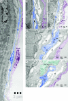
Telocytes in endocardium: electron microscope evidence
- PMID: 20716125
- PMCID: PMC3822573
- DOI: 10.1111/j.1582-4934.2010.01133.x
Telocytes in endocardium: electron microscope evidence
Abstract
The term TELOCYTES was very recently introduced, for replacing the name Interstitial Cajal-Like Cells (ICLC). In fact, telocytes are not really Cajal-like cells, they being different from all other interstitial cells by the presence of telopodes, which are cell-body prolongations, very thin (under the resolving power of light microscopy), extremely long (tens up to hundreds of micrometers), with a moniliform aspect (many dilations along), and having caveolae. The presence of telocytes in epicardium and myocardium was previously documented. We present here electron microscope images showing the existence of telocytes, with telopodes, at the level of mouse endocardium. Telocytes are located in the subendothelial layer of endocardium, and their telopodes are interposed in between the endocardial endothelium and the cardiomyocytes bundles. Some telopodes penetrate from the endocardium among the cardiomyocytes and surround them, eventually. Telopodes frequently establish close spatial relationships with myocardial blood capillaries and nerve endings. Because we may consider endocardium as a 'blood-heart barrier', or more exactly as a 'blood-myocardium barrier', telocytes might have an important role in such a barrier being the dominant cell population in subendothelial layer of endocardium.
© 2010 The Authors Journal compilation © 2010 Foundation for Cellular and Molecular Medicine/Blackwell Publishing Ltd.
Figures


References
-
- Covell JW. Tissue structure and ventricular wall mechanics. Circulation. 2008;118:699–701. - PubMed
-
- Anderson RH, Smerup M, Sanchez-Quintana D, et al. The three-dimensional arrangement of the myocytes in the ventricular walls. Clin Anat. 2009;22:64–76. - PubMed
-
- Zhou B, Pu WT. More than a cover: epicardium as a novel source of cardiac progenitor cells. Regen Med. 2008;3:633–5. - PubMed
-
- Habib M, Caspi O, Gepstein L. Human embryonic stem cells for cardiomyogenesis. J Mol Cell Cardiol. 2008;45:462–74. - PubMed
Publication types
MeSH terms
LinkOut - more resources
Full Text Sources

