

Histological grading of breast cancer on needle core biopsy: the role of immunohistochemical assessment of proliferation
- PMID: 20716163
- PMCID: PMC3564399
- DOI: 10.1111/j.1365-2559.2010.03620.x
Histological grading of breast cancer on needle core biopsy: the role of immunohistochemical assessment of proliferation
Abstract
Aims: Histological grade assessed on needle core biopsy (NCB) moderately concurs with the grade in the surgical excision specimen (SES) (kappa-values between 0.35 and 0.65). A major cause of the discrepancy is underestimation of mitoses in the NCB specimen. The aim was to determine the best method of assessing proliferation on NCB.
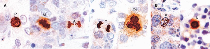
Methods and results: Proliferative activity of 101 invasive carcinomas of the breast on NCB and SES was assessed using mitotic counts on routine haematoxylin and eosin (H&E) sections and immunohistochemical markers Mib-1 and phosphorylated histone H3 (PPH3). H&E mitotic count in SES was considered as the gold standard. H&E mitotic count was found to be underestimated on NCB when compared with that in SES (P < 0.001), but no significant difference was detected between NCB and SES regarding Mib-1 (P = 0.13) or PPH3 (P = 0.073). Using receiver-operating characteristic curve, Mib-1 on NCB was found to agree with the gold standard significantly better than routine H&E on NCB.
Conclusions: Immunohistochemical markers in NCB showed better concordance with H&E mitotic count in SES (gold standard) than routine H&E mitotic count in NCB. Further refinement of cut-offs and scoring methods is needed.
Figures


Similar articles
-
Grading of breast cancer on needle core biopsy: does a reduction in mitotic count threshold improve agreement with grade on excised specimens?J Clin Pathol. 2014 Dec;67(12):1106-8. doi: 10.1136/jclinpath-2014-202294. Epub 2014 Sep 2. J Clin Pathol. 2014. PMID: 25185140
-
Federation Nationale des Centers de Lutte Contre le Cancer grading of soft tissue sarcomas on needle core biopsies using surrogate markers.Hum Pathol. 2016 Oct;56:147-54. doi: 10.1016/j.humpath.2016.06.008. Epub 2016 Jun 23. Hum Pathol. 2016. PMID: 27346575
-
Better see to better agree: phosphohistone H3 increases interobserver agreement in mitotic count for meningioma grading and imposes new specific thresholds.Neuro Oncol. 2015 May;17(5):663-9. doi: 10.1093/neuonc/nov002. Epub 2015 Feb 1. Neuro Oncol. 2015. PMID: 25646026 Free PMC article.
-
Correlation between MIB-1 and other proliferation markers: clinical implications of the MIB-1 cutoff value.Cancer. 2002 Apr 15;94(8):2151-9. doi: 10.1002/cncr.10458. Cancer. 2002. PMID: 12001111 Review.
-
Impact of molecular subtypes classification concordance between preoperative core needle biopsy and surgical specimen on early breast cancer management: Single-institution experience and review of published literature.Eur J Surg Oncol. 2017 Apr;43(4):642-648. doi: 10.1016/j.ejso.2016.10.025. Epub 2016 Nov 17. Eur J Surg Oncol. 2017. PMID: 27889196 Review.
Cited by
-
Photon Absorption Remote Sensing Imaging of Breast Needle Core Biopsies Is Diagnostically Equivalent to Gold Standard H&E Histologic Assessment.Curr Oncol. 2023 Nov 6;30(11):9760-9771. doi: 10.3390/curroncol30110708. Curr Oncol. 2023. PMID: 37999128 Free PMC article.
-
Does shear wave ultrasound independently predict axillary lymph node metastasis in women with invasive breast cancer?Breast Cancer Res Treat. 2014 Jan;143(1):153-7. doi: 10.1007/s10549-013-2747-z. Epub 2013 Dec 4. Breast Cancer Res Treat. 2014. PMID: 24305976 Free PMC article.
-
Can ki-67 play a role in prediction of breast cancer patients' response to neoadjuvant chemotherapy?Biomed Res Int. 2014;2014:628217. doi: 10.1155/2014/628217. Epub 2014 Mar 25. Biomed Res Int. 2014. PMID: 24783217 Free PMC article.
-
Long Non-Coding RNAs as Potential Diagnostic and Prognostic Biomarkers in Breast Cancer: Progress and Prospects.Front Oncol. 2021 Aug 30;11:710538. doi: 10.3389/fonc.2021.710538. eCollection 2021. Front Oncol. 2021. PMID: 34527584 Free PMC article. Review.
-
Evaluation of Ki67 expression across distinct categories of breast cancer specimens: a population-based study of matched surgical specimens, core needle biopsies and tissue microarrays.PLoS One. 2014 Nov 6;9(11):e112121. doi: 10.1371/journal.pone.0112121. eCollection 2014. PLoS One. 2014. PMID: 25375149 Free PMC article.
References
-
- Cahill RA, Walsh D, Landers RJ, Watson RG. Preoperative profiling of symptomatic breast cancer by diagnostic core biopsy. Ann. Surg. Oncol. 2006;13:45–51. - PubMed
-
- Puglisi F, Scalone S, Bazzocchi M, et al. Image-guided core breast biopsy: a suitable method for preoperative biological characterization of small (pT1) breast carcinomas. Cancer Lett. 1998;133:223–229. - PubMed
-
- Ogston KN, Miller ID, Payne S, et al. A new histological grading system to assess response of breast cancers to primary chemotherapy: prognostic significance and survival. Breast. 2003;12:320–327. - PubMed
-
- Elston CW, Ellis IO. Pathological prognostic factors in breast-cancer. 1. The value of histological grade in breast-cancer—experience from a large study with long-term follow-up. Histopathology. 1991;19:403–410. - PubMed
Publication types
MeSH terms
Substances
LinkOut - more resources
Full Text Sources
Medical
