

Characterization and functional analysis of seven flagellin genes in Rhizobium leguminosarum bv. viciae. Characterization of R. leguminosarum flagellins
- PMID: 20716375
- PMCID: PMC2936354
- DOI: 10.1186/1471-2180-10-219
Characterization and functional analysis of seven flagellin genes in Rhizobium leguminosarum bv. viciae. Characterization of R. leguminosarum flagellins
Abstract
Background: Rhizobium leguminosarum bv. viciae establishes symbiotic nitrogen fixing partnerships with plant species belonging to the Tribe Vicieae, which includes the genera Vicia, Lathyrus, Pisum and Lens. Motility and chemotaxis are important in the ecology of R. leguminosarum to provide a competitive advantage during the early steps of nodulation, but the mechanisms of motility and flagellar assembly remain poorly studied. This paper addresses the role of the seven flagellin genes in producing a functional flagellum.
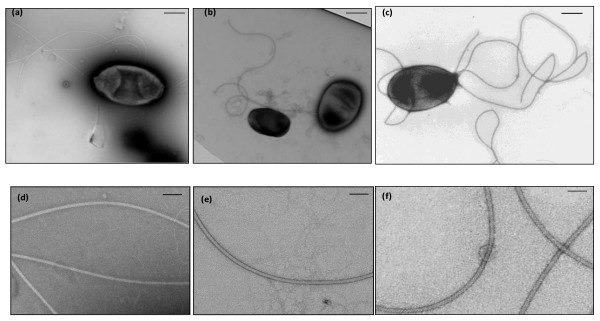
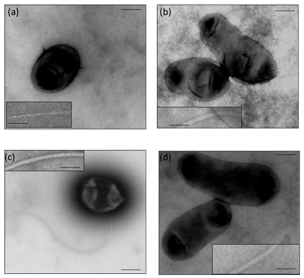
Results: R. leguminosarum strains 3841 and VF39SM have seven flagellin genes (flaA, flaB, flaC, flaD, flaE, flaH, and flaG), which are transcribed separately. The predicted flagellins of 3841 are highly similar or identical to the corresponding flagellins in VF39SM. flaA, flaB, flaC, and flaD are in tandem array and are located in the main flagellar gene cluster. flaH and flaG are located outside of the flagellar/motility region while flaE is plasmid-borne. Five flagellin subunits (FlaA, FlaB, FlaC, FlaE, and FlaG) are highly similar to each other, whereas FlaD and FlaH are more distantly related. All flagellins exhibit conserved amino acid residues at the N- and C-terminal ends and are variable in the central regions. Strain 3841 has 1-3 plain subpolar flagella while strain VF39SM exhibits 4-7 plain peritrichous flagella. Three flagellins (FlaA/B/C) and five flagellins (FlaA/B/C/E/G) were detected by mass spectrometry in the flagellar filaments of strains 3841 and VF39SM, respectively. Mutation of flaA resulted in non-motile VF39SM and extremely reduced motility in 3841. Individual mutations of flaB and flaC resulted in shorter flagellar filaments and consequently reduced swimming and swarming motility for both strains. Mutant VF39SM strains carrying individual mutations in flaD, flaE, flaH, and flaG were not significantly affected in motility and filament morphology. The flagellar filament and the motility of 3841 strains with mutations in flaD and flaG were not significantly affected while flaE and flaH mutants exhibited shortened filaments and reduced swimming motility.
Conclusion: The results obtained from this study demonstrate that FlaA, FlaB, and FlaC are major components of the flagellar filament while FlaD and FlaG are minor components for R. leguminosarum strains 3841 and VF39SM. We also observed differences between the two strains, wherein FlaE and FlaH appear to be minor components of the flagellar filaments in VF39SM but these flagellin subunits may play more important roles in 3841. This paper also demonstrates that the flagellins of 3841 and VF39SM are possibly glycosylated.
Figures


Similar articles
-
Regulation of flagellar, motility and chemotaxis genes in Rhizobium leguminosarum by the VisN/R-Rem cascade.Microbiology (Reading). 2010 Jun;156(Pt 6):1673-1685. doi: 10.1099/mic.0.035386-0. Epub 2010 Mar 4. Microbiology (Reading). 2010. PMID: 20203055
-
Multiple Flagellin Proteins Have Distinct and Synergistic Roles in Agrobacterium tumefaciens Motility.J Bacteriol. 2018 Nov 6;200(23):e00327-18. doi: 10.1128/JB.00327-18. Print 2018 Dec 1. J Bacteriol. 2018. PMID: 30201783 Free PMC article.
-
A Critical Region in the FlaA Flagellin Facilitates Filament Formation of the Vibrio cholerae Flagellum.J Bacteriol. 2018 Jul 10;200(15):e00029-18. doi: 10.1128/JB.00029-18. Print 2018 Aug 1. J Bacteriol. 2018. PMID: 29581407 Free PMC article.
-
The archaeabacterial flagellar filament: a bacterial propeller with a pilus-like structure.J Mol Microbiol Biotechnol. 2006;11(3-5):208-20. doi: 10.1159/000094055. J Mol Microbiol Biotechnol. 2006. PMID: 16983196 Review.
-
Transcription Regulation of Flagellins: A Structural Perspective.Biochemistry. 2025 Feb 18;64(4):770-781. doi: 10.1021/acs.biochem.4c00791. Epub 2025 Jan 28. Biochemistry. 2025. PMID: 39874281 Review.
Cited by
-
Swimming performance of Bradyrhizobium diazoefficiens is an emergent property of its two flagellar systems.Sci Rep. 2016 Apr 7;6:23841. doi: 10.1038/srep23841. Sci Rep. 2016. PMID: 27053439 Free PMC article.
-
Transcriptome profiling of a Rhizobium leguminosarum bv. trifolii rosR mutant reveals the role of the transcriptional regulator RosR in motility, synthesis of cell-surface components, and other cellular processes.BMC Genomics. 2015 Dec 29;16:1111. doi: 10.1186/s12864-015-2332-4. BMC Genomics. 2015. PMID: 26715155 Free PMC article.
-
Genome-wide analyses of Liberibacter species provides insights into evolution, phylogenetic relationships, and virulence factors.Mol Plant Pathol. 2020 May;21(5):716-731. doi: 10.1111/mpp.12925. Epub 2020 Feb 28. Mol Plant Pathol. 2020. PMID: 32108417 Free PMC article.
-
Tyrosine Nitration of Flagellins: a Response of Sinorhizobium meliloti to Nitrosative Stress.Appl Environ Microbiol. 2020 Dec 17;87(1):e02210-20. doi: 10.1128/AEM.02210-20. Print 2020 Dec 17. Appl Environ Microbiol. 2020. PMID: 33067191 Free PMC article.
-
Bacterial Flagellar Filament: A Supramolecular Multifunctional Nanostructure.Int J Mol Sci. 2021 Jul 14;22(14):7521. doi: 10.3390/ijms22147521. Int J Mol Sci. 2021. PMID: 34299141 Free PMC article. Review.
References
-
- Scharf B, Schuster-Wolff-Buhring H, Rachel R, Schmitt R. Mutational analysis of the Rhizobium lupini H13-3 and Sinorhizobium meliloti flagellin genes: importance of flagellin A for flagellar filament structure and transcriptional regulation. J Bacteriol. 2001;183:5334–5342. doi: 10.1128/JB.183.18.5334-5342.2001. - DOI - PMC - PubMed
Publication types
MeSH terms
Substances
LinkOut - more resources
Full Text Sources
