

Mutational analysis of an archaeal minichromosome maintenance protein exterior hairpin reveals critical residues for helicase activity and DNA binding
- PMID: 20716382
- PMCID: PMC2933578
- DOI: 10.1186/1471-2199-11-62
Mutational analysis of an archaeal minichromosome maintenance protein exterior hairpin reveals critical residues for helicase activity and DNA binding
Abstract
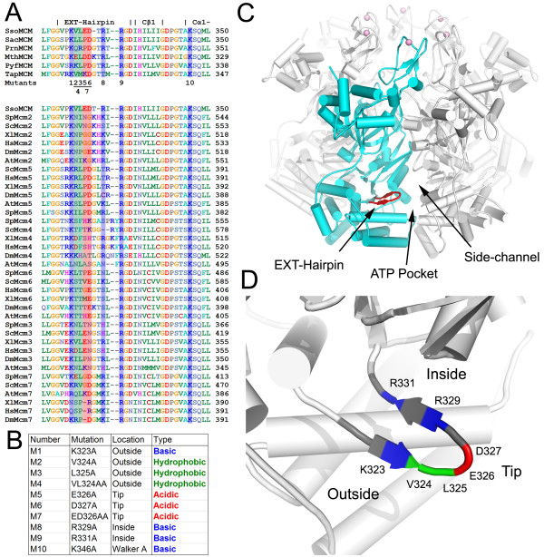
Background: The mini-chromosome maintenance protein (MCM) complex is an essential replicative helicase for DNA replication in Archaea and Eukaryotes. While the eukaryotic complex consists of six homologous proteins (MCM2-7), the archaeon Sulfolobus solfataricus has only one MCM protein (ssoMCM), six subunits of which form a homohexamer. We have recently reported a 4.35A crystal structure of the near full-length ssoMCM. The structure reveals a total of four beta-hairpins per subunit, three of which are located within the main channel or side channels of the ssoMCM hexamer model generated based on the symmetry of the N-terminal Methanothermobacter thermautotrophicus (mtMCM) structure. The fourth beta-hairpin, however, is located on the exterior of the hexamer, near the exit of the putative side channels and next to the ATP binding pocket.
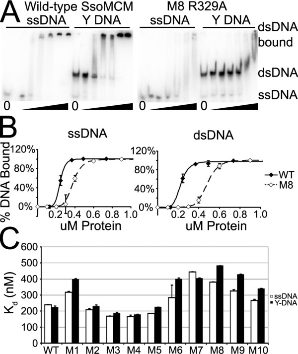
Results: In order to better understand this hairpin's role in DNA binding and helicase activity, we performed a detailed mutational and biochemical analysis of nine residues on this exterior beta-hairpin (EXT-hp). We examined the activities of the mutants related to their helicase function, including hexamerization, ATPase, DNA binding and helicase activities. The assays showed that some of the residues on this EXT-hp play a role for DNA binding as well as for helicase activity.
Conclusions: These results implicate several current theories regarding helicase activity by this critical hexameric enzyme. As the data suggest that EXT-hp is involved in DNA binding, the results reported here imply that the EXT-hp located near the exterior exit of the side channels may play a role in contacting DNA substrate in a manner that affects DNA unwinding.
Figures


Similar articles
-
Crystal structure of a near-full-length archaeal MCM: functional insights for an AAA+ hexameric helicase.Proc Natl Acad Sci U S A. 2008 Dec 23;105(51):20191-6. doi: 10.1073/pnas.0808037105. Epub 2008 Dec 10. Proc Natl Acad Sci U S A. 2008. PMID: 19073923 Free PMC article.
-
The Sulfolobus solfataricus GINS Complex Stimulates DNA Binding and Processive DNA Unwinding by Minichromosome Maintenance Helicase.J Bacteriol. 2015 Nov;197(21):3409-20. doi: 10.1128/JB.00496-15. Epub 2015 Aug 17. J Bacteriol. 2015. PMID: 26283767 Free PMC article.
-
Archaeal MCM has separable processivity, substrate choice and helicase domains.Nucleic Acids Res. 2007;35(3):988-98. doi: 10.1093/nar/gkl1117. Epub 2007 Jan 26. Nucleic Acids Res. 2007. PMID: 17259218 Free PMC article.
-
Unwinding the structure and function of the archaeal MCM helicase.Mol Microbiol. 2009 Apr;72(2):286-96. doi: 10.1111/j.1365-2958.2009.06663.x. Mol Microbiol. 2009. PMID: 19415794 Review.
-
Archaeal MCM Proteins as an Analog for the Eukaryotic Mcm2-7 Helicase to Reveal Essential Features of Structure and Function.Archaea. 2015 Oct 11;2015:305497. doi: 10.1155/2015/305497. eCollection 2015. Archaea. 2015. PMID: 26539061 Free PMC article. Review.
Cited by
-
The Helicase Activity of Hyperthermophilic Archaeal MCM is Enhanced at High Temperatures by Lysine Methylation.Front Microbiol. 2015 Nov 9;6:1247. doi: 10.3389/fmicb.2015.01247. eCollection 2015. Front Microbiol. 2015. PMID: 26617586 Free PMC article.
-
Molecular architecture of a multifunctional MCM complex.Nucleic Acids Res. 2012 Feb;40(3):1366-80. doi: 10.1093/nar/gkr831. Epub 2011 Oct 7. Nucleic Acids Res. 2012. PMID: 21984415 Free PMC article.
-
Steric exclusion and wrapping of the excluded DNA strand occurs along discrete external binding paths during MCM helicase unwinding.Nucleic Acids Res. 2011 Aug;39(15):6585-95. doi: 10.1093/nar/gkr345. Epub 2011 May 16. Nucleic Acids Res. 2011. PMID: 21576224 Free PMC article.
-
Cdc45 (cell division cycle protein 45) guards the gate of the Eukaryote Replisome helicase stabilizing leading strand engagement.Proc Natl Acad Sci U S A. 2015 Jan 20;112(3):E249-58. doi: 10.1073/pnas.1422003112. Epub 2015 Jan 5. Proc Natl Acad Sci U S A. 2015. PMID: 25561522 Free PMC article.
-
Mini-chromosome maintenance complexes form a filament to remodel DNA structure and topology.Nucleic Acids Res. 2013 Mar 1;41(5):3446-56. doi: 10.1093/nar/gkt022. Epub 2013 Jan 29. Nucleic Acids Res. 2013. PMID: 23361460 Free PMC article.
References
Publication types
MeSH terms
Substances
Grants and funding
LinkOut - more resources
Full Text Sources
Research Materials
Miscellaneous
