Reduced intra-amygdala activity to positively valenced faces in adolescent schizophrenia offspring
- PMID: 20716480
- PMCID: PMC3174012
- DOI: 10.1016/j.schres.2010.07.023
Reduced intra-amygdala activity to positively valenced faces in adolescent schizophrenia offspring
Abstract
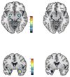
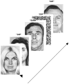
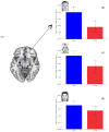
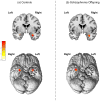
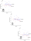
Studies suggest that the affective response is impaired in both schizophrenia and adolescent offspring of schizophrenia patients. Adolescent offspring of patients are developmentally vulnerable to impairments in several domains, including affective responding, yet the bases of these impairments and their relation to neuronal responses within the limbic system are poorly understood. The amygdala is the central region devoted to the processing of emotional valence and its sub-nuclei including the baso-lateral and centro-medial are organized in a relative hierarchy of affective processing. Outputs from the centro-medial nucleus converge on regions involved in the autonomous regulation of behavior, and outputs from the baso-lateral nucleus modulate the response of reward processing regions. Here using fMRI we assessed the intra-amygdala response to positive, negative, and neutral valenced faces in a group of controls (with no family history of psychosis) and offspring of schizophrenia parents (n=44 subjects in total). Subjects performed an affective continuous performance task during which they continually appraised whether the affect signaled by a face on a given trial was the same or different from the previous trial (regardless of facial identity). Relative to controls, offspring showed reduced activity in the left centro-medial nucleus to positively (but not negatively or neutral) valenced faces. These results were independent of behavioral/cognitive performance (equal across groups) suggesting that an impaired affective substrate in the intra-amygdala response may lie at the core of deficits of social behavior that have been documented in this population.
Copyright © 2010 Elsevier B.V. All rights reserved.
Conflict of interest statement
Figures

Similar articles
-
Amygdala and nucleus accumbens activation to emotional facial expressions in children and adolescents at risk for major depression.Am J Psychiatry. 2008 Jan;165(1):90-8. doi: 10.1176/appi.ajp.2007.06111917. Epub 2007 Nov 6. Am J Psychiatry. 2008. PMID: 17986682
-
Evidence for altered amygdala activation in schizophrenia in an adaptive emotion recognition task.Psychiatry Res. 2014 Mar 30;221(3):195-203. doi: 10.1016/j.pscychresns.2013.12.001. Epub 2013 Dec 10. Psychiatry Res. 2014. PMID: 24434194
-
Amygdala responses to positively and negatively valenced baby faces in healthy female volunteers: influences of individual differences in harm avoidance.Brain Res. 2009 Nov 3;1296:94-103. doi: 10.1016/j.brainres.2009.08.010. Epub 2009 Aug 11. Brain Res. 2009. PMID: 19679112
-
ADHD and schizophrenia phenomenology: visual scanpaths to emotional faces as a potential psychophysiological marker?Neurosci Biobehav Rev. 2006;30(5):651-65. doi: 10.1016/j.neubiorev.2005.11.004. Epub 2006 Feb 7. Neurosci Biobehav Rev. 2006. PMID: 16466794 Review.
-
Emotional face processing in schizophrenia.Curr Opin Psychiatry. 2009 Mar;22(2):140-6. doi: 10.1097/YCO.0b013e328324f895. Curr Opin Psychiatry. 2009. PMID: 19553867 Review.
Cited by
-
Bidirectional communication between amygdala and fusiform gyrus during facial recognition.Neuroimage. 2011 Jun 15;56(4):2348-55. doi: 10.1016/j.neuroimage.2011.03.072. Epub 2011 Apr 8. Neuroimage. 2011. PMID: 21497657 Free PMC article.
-
MET and AKT genetic influence on facial emotion perception.PLoS One. 2012;7(4):e36143. doi: 10.1371/journal.pone.0036143. Epub 2012 Apr 27. PLoS One. 2012. PMID: 22558359 Free PMC article.
-
Amygdalar and Hippocampal Morphometry Abnormalities in First-Episode Schizophrenia Using Deformation-Based Shape Analysis.Front Psychiatry. 2020 Jul 17;11:677. doi: 10.3389/fpsyt.2020.00677. eCollection 2020. Front Psychiatry. 2020. PMID: 32765318 Free PMC article.
-
Neurophysiological indices of face processing in people with psychosis and their siblings: An event-related potential study.Eur J Neurosci. 2024 Apr;59(8):1863-1876. doi: 10.1111/ejn.16034. Epub 2023 May 23. Eur J Neurosci. 2024. PMID: 37160716 Free PMC article.
-
Prediction errors and valence: From single units to multidimensional encoding in the amygdala.Behav Brain Res. 2021 Apr 23;404:113176. doi: 10.1016/j.bbr.2021.113176. Epub 2021 Feb 14. Behav Brain Res. 2021. PMID: 33596433 Free PMC article. Review.
References
-
- Adolphs R, Baron-Cohen S, Tranel D. Impaired recognition of social emotions following amygdala damage. J Cogn Neurosci. 2002;14:1264–1274. - PubMed
-
- Aleman A, Kahn RS. Strange feelings: do amygdala abnormalities dysregulate the emotional brain in schizophrenia? Prog Neurobiol. 2005;77:283–298. - PubMed
-
- Amaral DG, Behniea H, Kelly JL. Topographic organization of projections from the amygdala to the visual cortex in the macaque monkey. Neuroscience. 2003a;118:1099–1120. - PubMed
-
- Amaral DG, Capitanio JP, Jourdain M, Mason WA, Mendoza SP, Prather M. The amygdala: is it an essential component of the neural network for social cognition? Neuropsychologia. 2003b;41:235–240. - PubMed
-
- Amunts K, Kedo O, Kindler M, Pieperhoff P, Mohlberg H, Shah NJ, Habel U, Schneider F, Zilles K. Cytoarchitectonic mapping of the human amygdala, hippocampal region and entorhinal cortex: intersubject variability and probability maps. Anat Embryol (Berl) 2005;210:343–352. - PubMed
Publication types
MeSH terms
Grants and funding
LinkOut - more resources
Full Text Sources
Medical

