. 2010 Dec;38(22):8269-76.
doi: 10.1093/nar/gkq720. Epub 2010 Aug 16.

Autonomous zinc-finger nuclease pairs for targeted chromosomal deletion

Affiliations

Autonomous zinc-finger nuclease pairs for targeted chromosomal deletion

Cem Söllü et al. Nucleic Acids Res. 2010 Dec.

Abstract

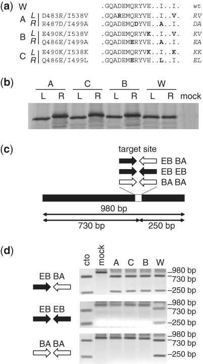
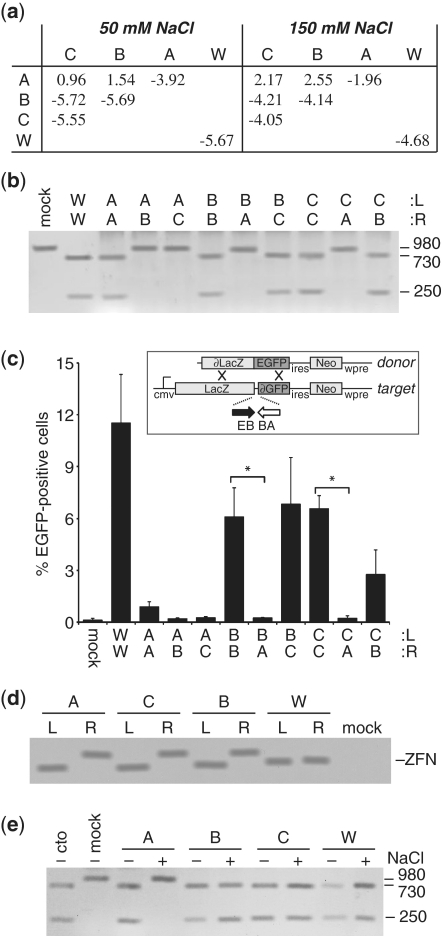
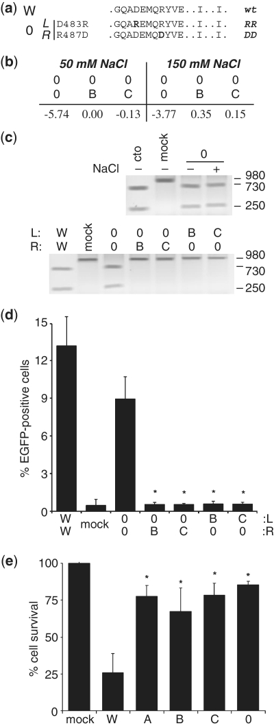
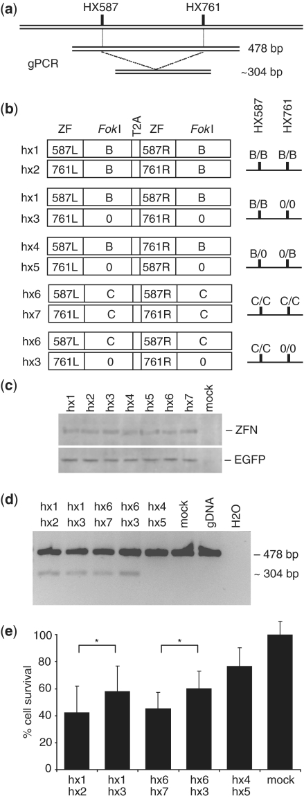
Zinc-finger nucleases (ZFNs) have been successfully used for rational genome engineering in a variety of cell types and organisms. ZFNs consist of a non-specific FokI endonuclease domain and a specific zinc-finger DNA-binding domain. Because the catalytic domain must dimerize to become active, two ZFN subunits are typically assembled at the cleavage site. The generation of obligate heterodimeric ZFNs was shown to significantly reduce ZFN-associated cytotoxicity in single-site genome editing strategies. To further expand the application range of ZFNs, we employed a combination of in silico protein modeling, in vitro cleavage assays, and in vivo recombination assays to identify autonomous ZFN pairs that lack cross-reactivity between each other. In the context of ZFNs designed to recognize two adjacent sites in the human HOXB13 locus, we demonstrate that two autonomous ZFN pairs can be directed simultaneously to two different sites to induce a chromosomal deletion in ∼ 10% of alleles. Notably, the autonomous ZFN pair induced a targeted chromosomal deletion with the same efficacy as previously published obligate heterodimeric ZFNs but with significantly less toxicity. These results demonstrate that autonomous ZFNs will prove useful in targeted genome engineering approaches wherever an application requires the expression of two distinct ZFN pairs.


Figures

Figure 1.
Figure 1.
In vitro cleavage assay with obligate heterodimeric ZFNs. (a) Sequence of the FokI variants. The sequence is indicated for FokI positions 480–490 (helix α4), 499 and 538, and is shown for wild-type (W) and variants (A, B, C). The two-letter code on the right defines the corresponding point mutations in each subunit. (b) In vitro translated ZFNs. ZFNs were expressed in the presence of 35S-met, separated by SDS–PAGE and exposed to X-ray film. L, left ZFN subunit EB2-N; R, right ZFN subunit BA1-N. (c) Schematic representation of in vitro cleavage assay. A linear DNA substrate containing the combined target site for ZFN subunits EB2-N and BA1-N (EB/BA) or inverted repeat target sites for subunits EB2-N (EB/EB) or BA1-N (BA/BA), respectively, are cleaved into different size products. (d) Analysis of heterodimeric or homodimeric cleavage reactions. A linear DNA substrate was incubated with in vitro translated ZFNs and the extent of cleavage analyzed by agarose gel electrophoresis. Control reactions were incubations with in vitro translated EGFP (mock) or AgeI (cto).
Figure 2.
Figure 2.
Comprehensive analysis of obligate heterodimeric ZFNs. (a) In silico computation of dimerization energy. The interaction energy of two variant ZFN subunits was calculated using FoldX based on structural models created with DeepView. The table indicates the average of the two calculated values (in kJ/mol) obtained when computing left-right versus right–left. (b) In vitro cleavage assay. A linear DNA substrate containing the heterodimeric target site for subunits EB2-N (L) and BA1-N (R) was incubated with in vitro translated ZFN variants in different combinations and the extent of cleavage analyzed by agarose gel electrophoresis. Control reactions were incubations with in vitro translated EGFP (mock) or wild-type ZFN (W). (c) Chromosomal recombination assay. Cell line 293EBBA contains an integrated mutated EGFP target locus (∂EGFP) with an adjacent heterodimeric target site for ZFN subunits EB2-N and BA1-N (EB/BA). After transfection with the various ZFN expression vectors and a donor plasmid, the percentage of EGFP-positive cells was assessed by flow cytometry. An expression vector encoding a mutated nuclease (mock) was included as a control to indicate the level of non-stimulated gene targeting. A statistically significant (P < 0.005) decrease in gene targeting between homologous and heterologous ZFN subunit combinations is indicated by asterisks. (d) ZFN expression levels. Transfected HEK293T cells were harvested after 30 h and lysates probed with an antibody against the HA tag. ‘mock’ indicates transfection with an EGFP-expression vector. (e) Sensitivity to high salt concentrations. The linear DNA target was incubated with the in vitro translated variant ZFN pairs and the extent of cleavage analyzed by agarose gel electrophoresis. (+) or (–) indicates the presence or absence of an additional 100 mM NaCl in the reaction. Control reactions were incubations with in vitro translated EGFP (mock) or AgeI (cto). A, B, C, and W denote FokI variants as defined in Figure 1a.
Figure 3.
Figure 3.
Identification of autonomous ZFN variants. (a) Sequence of the FokI variant pair 0. The sequence is indicated for FokI positions 480–490 (helix α4), 499 and 538. The two-letter code on the right defines the corresponding point mutation in each subunit. (b) In silico computation of dimerization energy. The table indicates the average of the two calculated values (in kJ/mol) obtained when computing left–right versus right–left. (c) In vitro cleavage assays. A linear DNA substrate was incubated with in vitro translated ZFNs and the extent of cleavage analyzed by agarose gel electrophoresis. Top: the linear DNA target was incubated with variant ZFN pair 0. (+) or (–) indicates the presence or absence of an additional 100 mM NaCl in the reaction. Bottom: subunits of variant 0 were combined with the respective subunits of variants B and C. Control reactions were incubations with in vitro translated EGFP (mock), wild-type ZFN (W) or AgeI (cto). (d) Chromosomal recombination assay. Target cell line 293EBBA was transfected as in (Figure 2c) with combinations of the various ZFN expression vectors. The columns designate the percentage of EGFP-positive cells 5 days after transfection, as determined by flow cytometry. An expression vector encoding a mutated nuclease (mock) was included as a negative control. A statistically significant (P < 0.002) decrease in gene targeting between homologous and heterologous ZFN subunit combinations is indicated by asterisks. (e) Quantitative cytotoxicity assay. HEK293 cells were transfected as described in (d). The columns represent the fraction of DsRed-Express-positive cells at day 5 as compared to the fraction at Day 2 after transfection, and are shown relative to transfection with an expression vector encoding a mutated nuclease (mock). A statistically significant increase in cell survival as compared to wild-type ZFN (W) is indicated by (*P < 0.01). A, B, C, 0 and W denote FokI variants.
Figure 4.
Figure 4.
Targeted chromosomal deletion in human HOXB13 locus. (a) Diagram of genomic PCR strategy. ZFN target sites (HX587 and HX761) and possible PCR products are indicated. (b) Schematic representation of HOXB13-specific ZFN expression constructs. Each expression plasmid encodes two ZFN subunits, which are separated by a sequence encoding the T2A autoproteinase. The plasmid names are shown on the left, the specificity of the DNA-binding domain and the respective FokI variant is indicated in the boxes. The FokI configuration of the five tested combinations at the two target sites is shown on the right. (c) ZFN expression levels. Lysates of HEK293T, which were co-transfected with ZFN expression vectors and pEGFP, were probed with antibodies against the FLAG tag and EGFP. Mock indicates transfection with empty vectors. (d) Targeted chromosomal deletion. HEK293T were transfected with pEGFP and the indicated plasmids. PCR was performed on extracted genomic DNA and fragments separated by agarose gel electrophoresis. The size of the fragments is indicated on the right. Mock indicates transfection with a mutant nuclease and gDNA denotes a non-transfected control sample. (e) Quantitative cytotoxicity assay. HEK293T cells were transfected as described in (d). The columns represent the average fraction of EGFP-positive cells at Day 5 as compared to the fraction at Day 2 after transfection, and are shown relative to transfection with an expression vector encoding a mutated nuclease (mock). A statistically significant increase in cell survival between combinations hx1/hx2 and hx1/hx3 or hx6/hx7 and hx6/hx3 is indicated by (*P < 0.05). B, C and 0 denote FokI variants.

Similar articles

Cited by

References

    1. Cathomen T, Schambach A. Zinc positive: tailored genome engineering meets reprogramming. Gene Ther. 2010;17:1–3. - PubMed
    1. Cathomen T, Joung KJ. Zinc-finger nucleases: the next generation emerges. Mol. Ther. 2008;16:1200–1207. - PubMed
    1. Carroll D. Progress and prospects: zinc-finger nucleases as gene therapy agents. Gene Ther. 2008;15:1463–1468. - PMC - PubMed
    1. Kim YG, Cha J, Chandrasegaran S. Hybrid restriction enzymes: zinc finger fusions to Fok I cleavage domain. Proc. Natl Acad. Sci. USA. 1996;93:1156–1160. - PMC - PubMed
    1. Rebar EJ, Pabo CO. Zinc finger phage: affinity selection of fingers with new DNA-binding specificities. Science. 1994;263:671–673. - PubMed

Publication types

MeSH terms

Substances