. 2010 Dec;38(22):8061-71.
doi: 10.1093/nar/gkq717. Epub 2010 Aug 16.

AUF1 p42 isoform selectively controls both steady-state and PGE2-induced FGF9 mRNA decay

Affiliations

AUF1 p42 isoform selectively controls both steady-state and PGE2-induced FGF9 mRNA decay

Tsung-Ming Chen et al. Nucleic Acids Res. 2010 Dec.

Abstract

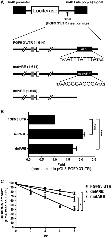
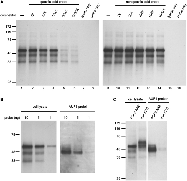
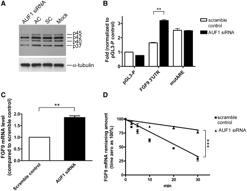
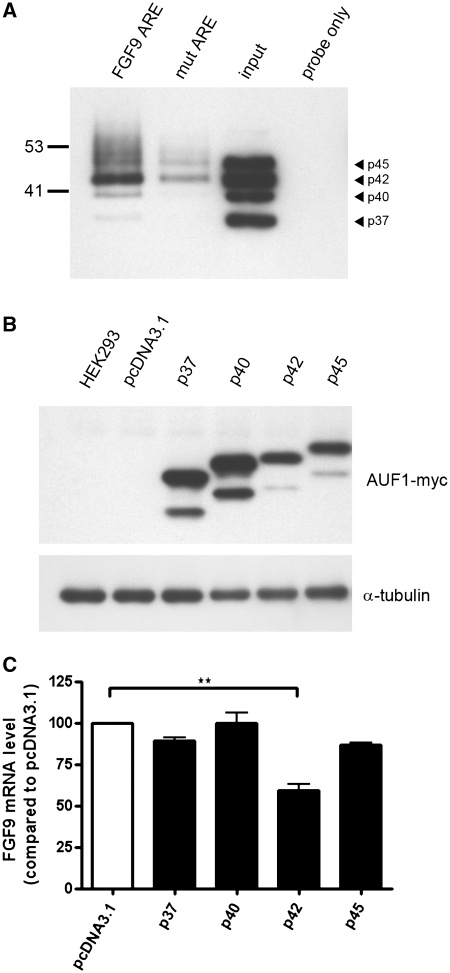
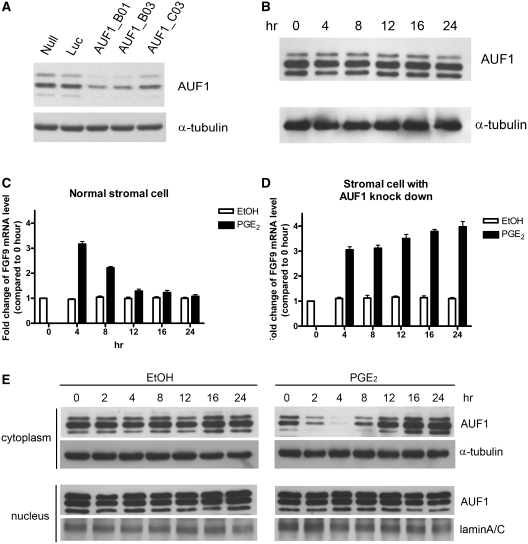
Fibroblast growth factor 9 (FGF9) is an autocrine/paracrine growth factor that plays vital roles in many physiologic processes including embryonic development. Aberrant expression of FGF9 causes human diseases and thus it highlights the importance of controlling FGF9 expression; however, the mechanism responsible for regulation of FGF9 expression is largely unknown. Here, we show the crucial role of an AU-rich element (ARE) in FGF9 3'-untranslated region (UTR) on controlling FGF9 expression. Our data demonstrated that AUF1 binds to this ARE to regulate FGF9 mRNA stability. Overexpression of each isoform of AUF1 (p37, p40, p42 and p45) showed that only the p42 isoform reduced the steady-state FGF9 mRNA. Also, knockdown of p42(AUF1) prolonged the half-life of FGF9 mRNA. The induction of FGF9 mRNA in prostaglandin (PG) E(2)-treated human endometrial stromal cells was accompanied with declined cytoplasmic AUF1. Nevertheless, ablation of AUF1 led to sustained elevation of FGF9 expression in these cells. Our study demonstrated that p42(AUF1) regulates both steady-state and PGE(2)-induced FGF9 mRNA stability through ARE-mediated mRNA degradation. Since almost half of the FGF family members are ARE-containing genes, our findings also suggest that ARE-mediated mRNA decay is a common pathway to control FGFs expression, and it represents a novel RNA regulon to coordinate FGFs homeostasis in various physiological conditions.


Figures

Figure 1.
Figure 1.
Sequence features of the human FGF9 3′-UTR. (A) The sequence corresponding to the 3′-UTR of FGF9 is depicted. The initial TGA nucleotide sequence (−3 to −1) corresponds to the translation stop codon. The GA and TG microsatellite motifs are marked in italic type. The ARE sequence (ATTTATTTA) is marked in bold and the underlines mark the poly-A signals (AATAAA). (B) Cross-species alignment of mouse, rat, human, cow, dog, swine and bird showed an overall 79% identity in the AU-rich element region.
Figure 2.
Figure 2.
FGF9 ARE decreases gene expression in HEK293 cells. (A) Diagrams showing the firefly luciferase reporter gene construct (top diagram), full-length FGF9 3′-UTR cloned into the 3′-end of the firefly luciferase reporter gene (second diagram), and constructs containing site-direct mutation of ARE (mutARE) (third diagram) and deletion of ARE (delARE) (fourth diagram). Empty and gray squares represent TG and GA dinucleotide repeats, respectively. The black square represents ARE motif. (B) Reporter gene activity shown as relative luciferase activity (fold) normalized to FGF9 3′-UTR. (C) Effect of FGF9 ARE on mRNA stability detected by real-time RT-qPCR. FGF9 3′-UTR (filled square), delARE (Filled triangle) or mutARE (filled inverted triangle) was transfected into HEK293 cells, which were treated with Act D (5 μg/ml) to inhibit transcription of the luciferase reporter gene. Results are normalized to time zero for each construct. All data are shown as mean ± SEM of 4–6 independent experiments. *P < 0.05; ***P < 0.001.
Figure 3.
Figure 3.
AUF1 specifically binds to FGF9 3′-UTR ARE. (A) UV cross-linking to biotin-labeled riboprobe from whole-cell lysate of HEK293 cells showed three protein–RNA complexes. Competition assays were performed by adding extra non-biotin-label (cold) riboprobe as competitor with labeled probe. Left panel, specific cold probe; right panel, non-specific cold probe. (B) Dose-dependence of proteins in cell lysate (left panel) and AUF1 (right panel) binding to FGF9 ARE. (C) UV cross-linking in cell lysate (left panel) and AUF1 protein (right panel) showing linkage to proteins in the wild-type FGF9 ARE but not to the mutant ARE.
Figure 4.
Figure 4.
AUF1 controls FGF9 mRNA stability. (A) Western blots showing AUF1-specific siRNA, but not the scramble control (SC) or AUF1-specific siRNA control (AC), knocks down AUF1 protein. Mock is HEK293 cells treated with transfecting reagent only and α-tubulin was used as a control. (B) Luciferase reporter activities in empty vector (pGL3-P), full length (FGF9 3′-UTR), and mutant (mutARE) after AUF1 or scramble siRNA knockdown were shown. (C) Endogenous FGF9 mRNA levels in HEK293 cells following AUF1 or scramble siRNA knockdown. (D) Real-time RT-qPCR measurements showing the half-life of endogenous FGF9 mRNA with or without siRNA knockdown. Results are normalized to time zero for each construct. All data are shown as mean ± SEM of 4–6 independent experiments. **P < 0.01; ***P < 0.001.
Figure 5.
Figure 5.
AUF1 p42 isoform selectively downregulates FGF9 mRNA expression. (A) Pull-down assay showing that p42AUF1 is the major isoform pulled down by biotin-labeled FGF9 ARE probe. Arrowhead indicates the position of each AUF1 isoform. Note that the mutant is markedly weaker than the wild type. Input and probe only indicate positive and negative controls. (B) Western blots using anti-Myc antibody showing the expression of indicated recombinant AUF1 proteins. HEK293 and pcDNA3.1 refer to cells without transfection and cells transfected with empty vector, respectively. (C) Endogenous FGF9 mRNA levels relative to control (pcDNA3.1) in cells overexpressing the AUF1 isoforms p37, p40, p42 and p45. **P < 0.01.
Figure 6.
Figure 6.
AUF1 knockdown sustains the PGE2-medicated induction of FGF9 mRNA in human primary endometrial stromal cells. (A) Western blots showing successful AUF1 knockdown by AUF1-specific shRNA (B01, B03 and C03), but not the Luc control shRNA. The Null indicates cells without shRNA treatment. (B) Western blots showing AUF1 protein expression in whole-cell lysates isolated from stromal cells after PGE2 treatment. (C) FGF9 mRNA levels measured at indicated times after PGE2 treatment in stromal cells. Ethanol (EtOH) was used as control treatment. (D) Experiment similar to that shown in C, but with AUF1 knockdown by AUF1_B03 shRNA. All data are shown as mean ± SEM of 4–6 independent experiments. *P < 0.05; **P < 0.01; ***P < 0.001. (E) Western blots showing the distribution pattern of AUF1 protein in stromal cells after PGE2 treatment. α-Tubulin and LaminA/C used as loading controls for proteins isolated from cytoplasmic and nuclear fractions, respectively.

References

    1. Beenken A, Mohammadi M. The FGF family: biology, pathophysiology and therapy. Nat. Rev. Drug Discov. 2009;8:235–253. - PMC - PubMed
    1. Goldfarb M. Functions of fibroblast growth factors in vertebrate development. Cytokine Growth Factor Rev. 1996;7:311–325. - PubMed
    1. Gerwins P, Skoldenberg E, Claesson-Welsh L. Function of fibroblast growth factors and vascular endothelial growth factors and their receptors in angiogenesis. Crit. Rev. Oncol. Hematol. 2000;34:185–194. - PubMed
    1. Allouche M, Bikfalvi A. The role of fibroblast growth factor-2 (FGF-2) in hematopoiesis. Prog. Growth Factor Res. 1995;6:35–48. - PubMed
    1. Galzie Z, Kinsella AR, Smith JA. Fibroblast growth factors and their receptors. Biochem. Cell Biol. 1997;75:669–685. - PubMed

Publication types

MeSH terms