. 2010 Nov 12;285(46):36049-59.
doi: 10.1074/jbc.M110.110189. Epub 2010 Aug 17.

A major fraction of glycosphingolipids in model and cellular cholesterol-containing membranes is undetectable by their binding proteins

Affiliations

A major fraction of glycosphingolipids in model and cellular cholesterol-containing membranes is undetectable by their binding proteins

Radhia Mahfoud et al. J Biol Chem. .

Abstract

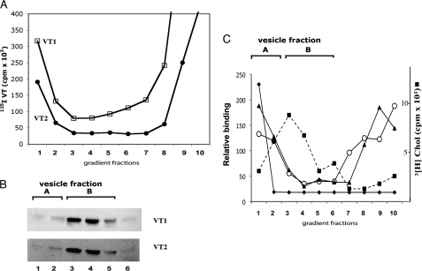
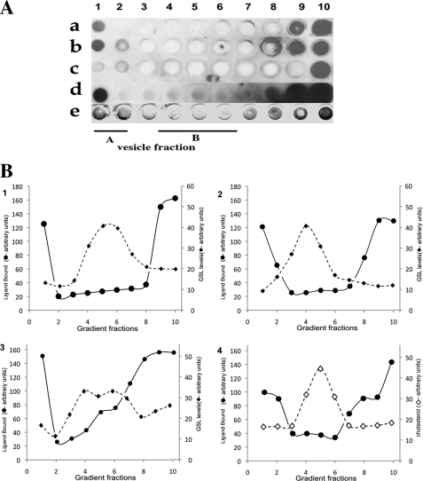
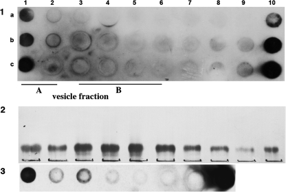
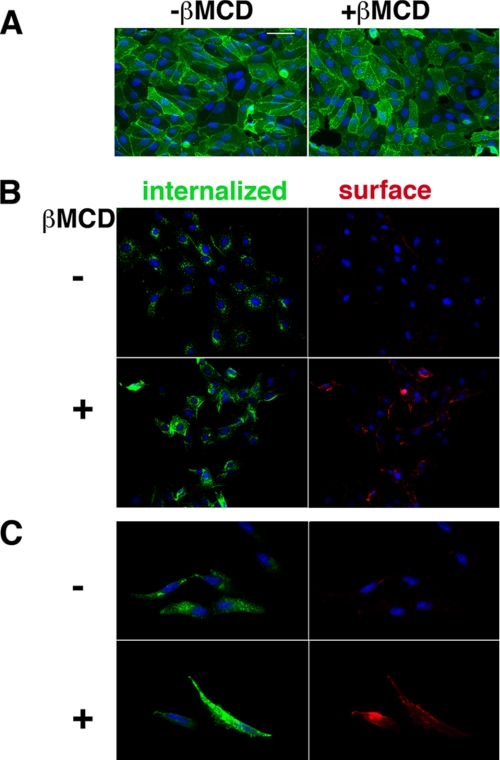
Glycosphingolipids (GSLs) accumulate in cholesterol-enriched cell membrane domains and provide receptors for protein ligands. Lipid-based "aglycone" interactions can influence GSL carbohydrate epitope presentation. To evaluate this relationship, Verotoxin binding its receptor GSL, globotriaosyl ceramide (Gb(3)), was analyzed in simple GSL/cholesterol, detergent-resistant membrane vesicles by equilibrium density gradient centrifugation. Vesicles separated into two Gb(3/)cholesterol-containing populations. The lighter, minor fraction (<5% total GSL), bound VT1, VT2, IgG/IgM mAb anti-Gb(3), HIVgp120 or Bandeiraea simplicifolia lectin. Only IgM anti-Gb(3), more tolerant of carbohydrate modification, bound both vesicle fractions. Post-embedding cryo-immuno-EM confirmed these results. This appears to be a general GSL-cholesterol property, because similar receptor-inactive vesicles were separated for other GSL-protein ligand systems; cholera toxin (CTx)-GM1, HIVgp120-galactosyl ceramide/sulfatide. Inclusion of galactosyl or glucosyl ceramide (GalCer and GlcCer) rendered VT1-unreactive Gb(3)/cholesterol vesicles, VT1-reactive. We found GalCer and GlcCer bind Gb(3), suggesting GSL-GSL interaction can counter cholesterol masking of Gb(3). The similar separation of Vero cell membrane-derived vesicles into minor "binding," and major "non-binding" fractions when probed with VT1, CTx, or anti-SSEA4 (a human GSL stem cell marker), demonstrates potential physiological relevance. Cell membrane GSL masking was cholesterol- and actin-dependent. Cholesterol depletion of Vero and HeLa cells enabled differential VT1B subunit labeling of "available" and "cholesterol-masked" plasma membrane Gb(3) pools by fluorescence microscopy. Thus, the model GSL/cholesterol vesicle studies predicted two distinct membrane GSL formats, which were demonstrated within the plasma membrane of cultured cells. Cholesterol masking of most cell membrane GSLs may impinge many GSL receptor functions.


Figures

FIGURE 1.
FIGURE 1.
Gb3/cholesterol vesicles can be separated into minor VT1/VT2-binding and major non-binding fractions. A, 125I-VT1 (□) and 125I-VT2 (●), added to vesicles before gradient centrifugation. B, Gb3 in gradient fractions was extracted and detected by VT1/VT2-TLC overlay. C, densitometry of immunodetected exogenous VT1 (♦) or VT2 (▴) binding to gradient-separated Gb3/cholesterol vesicle, VT1 distribution (○) in gradient fractions when added before separation. The distribution of [3H]cholesterol (■) is shown.
FIGURE 2.
FIGURE 2.
Other Gb3-binding proteins show a similar vesicle binding profile. The mouse IgG mAb anti-Gb3 (clone BGR23) was compared with the rat IgM mAb anti-Gb3 (clone 38.13) and B. simplicifolia for binding sucrose gradient-separated Gb3/cholesterol vesicles. Panel i: lanes a–c and d–f, increasing aliquots of the gradient-separated Gb3 vesicles stained with the BGR23 and 38.13, respectively. Panel ii: binding of VT1 (lane a), B. simplicifolia lectin (lane b), and 38.13 (lane c) to sucrose gradient-separated Gb3/cholesterol vesicles.
FIGURE 3.
FIGURE 3.
Post-embedding cryo-immuno-EM of Gb3/cholesterol vesicles confirms VT1 vesicle binding profile. VT1 immunogold labeling of unfractionated Gb3/cholesterol vesicles (A–D), sucrose gradient vesicle fraction A (F), and vesicle fraction B (E). IgM mAb anti-Gb3 labeling of vesicle fraction B is in panel G. Because the VT1-antibody-gold or IgM anti-Gb3-gold complex is at least 50 Å, surface binding is conclusive only when gold is external to the vesicle limiting membrane. In unseparated samples, larger vesicles (A and B) are VT1-labeled internally only, whereas VT1 labels the both bilayer leaflets of smaller vesicles (C and D, arrows). Inner membrane VT1 labeling of gradient separated vesicle fraction B is seen, whereas the outer membrane (E, arrows) is unlabeled. The smaller vesicles found in fraction A show outer membrane VT1 labeling (F, arrow). Rat IgM anti-Gb3 outer membrane labeling of vesicle fraction B is shown in G (arrows). Bar = 100 nm. Controls in which VT1 was omitted or rat IgM isotype control used showed no labeling.
FIGURE 4.
FIGURE 4.
R5 HIV-1 gp120 binding is similarly restricted to a minor fraction of GSL/cholesterol vesicles. GSL/cholesterol vesicles were mixed with gp120, separated by sucrose gradient, and immobilized on nitrocellulose for bound gp120 immunodetection. A, gp120 binding to Gb3 (a), GalCer (b), Gb4 (c), and SGC gradient fractions (d); e, VT1 binding to Gb3 sucrose gradient-separated vesicles. B, ligand-GSL binding in A was compared by densitometry (●) to gradient fraction GSL content (♦). GSLs were extracted, separated by TLC, and quantitated by densitometry of the orcinol stain. 1, gp120 and Gb3; 2, gp120 and GalCer; 3, gp120 and SGC; 4, VT1 binding Gb3 (●) compared with cholesterol (♢) distribution by FeCl3 detection after TLC.
FIGURE 5.
FIGURE 5.
Cholera toxin also binds a minor subfraction of GM1/cholesterol vesicles. Cholera toxin was added above GM1/cholestrol vesicle constructs and vesicles separated on a discontinuous sucrose gradient. After separation, fractions (1 (top)-10) were tested for cholera toxin content (panel 1). Cholera toxin binding is only found in fraction 1 (vesicle fraction A). GM1 extracted from gradient fractions was detected by CTxB TLC overlay (panel 2). GM1 is distributed in the gradient but accumulates in fractions 3, 4, 5, and 6 (vesicle fraction B). Anti-GM1 was included in a similar GM1/cholesterol vesicle gradient (panel 3) and immunodetected in the separated fractions.
FIGURE 6.
FIGURE 6.
Galactosyl or glucosyl ceramide can unmask Gb3/cholesterol for VT1 binding, and bind Gb3 directly. A, Gb3/cholesterol vesicles were separated in a VT1-containing sucrose gradient and vesicle-bound VT1 detected by immunoblot and compared with Gb3 distribution. Panels a–c: upper section, VT1-TLC overlay to detect Gb3 in GSL extract of gradient fractions: left-most lane, Gb3 standard; lower section: immunodetection of vesicle-bound VT1 in gradient fractions. a, Gb3/cholesterol vesicles; b, GalCer (non-hydroxy fatty acid)/Gb3/cholesterol vesicles; c, GalCer (hydroxy fatty acid)/Gb3/cholesterol vesicles. Vesicle fractions A and B are indicated as bars above the panels. B, GSLs were screened for Gb3 binding. Purified lipids (GalCer, GlcCer, adamantyl-GalCer, SGC, LacCer, lysoGalCer, cholesterol, Gb4, and Gb3) were spotted on TLC and detected by orcinol (a). Similar plates were incubated ± “aqueous” Gb3 and then tested for VT1 binding (b and c). C, GalCer, GlcCer, and LacCer were compared for effect on Gb3/cholesterol vesicle binding in a VT1-containing sucrose gradient. Vesicle-bound VT1 was detected by immunoblot. a, GalCer (non-hydroxy fatty acid)Gb3/cholesterol vesicles; b, GalCer (hydroxy fatty acid)/Gb3/cholesterol vesicles; c, GlcCer/Gb3/cholesterol vesicles; d, LacCer/Gb3/cholesterol vesicles. Vesicle fractions A and B are indicated as bars above panel C.
FIGURE 7.
FIGURE 7.
Cell membrane-derived vesicles show similar, cholesterol dependent, minor ligand-bound, and major ligand-unbound GSL fractions. The Triton extracts of cell membranes prepared from control Vero cells or cells grown in P4 to deplete Gb3, were separated on a VT1 containing sucrose gradient. Control cell membranes were also extracted ± βMCD or treated ± latrunculin prior to detergent treatment. Vesicle-bound VT1 was detected in dot-blotted gradient fractions, and total VT1 by Western blot of gradient fractions. mAb anti-SSEA4 (globoseries embryonic GSL antigen) binding to gradient-separated Vero cell vesicles was assessed. In A: a, immunoblot detecting VT1 binding to control Vero cell membrane vesicles; b, VT1 binding to membrane vesicles from P4-treated (GSL-depleted) cells; c, phospholipids within the lipid extract of gradient fractions separated by TLC, were detected by iodine staining (upper band, PE; lower, PC); d, simultaneous detection of Gb3 (*) and GM1 (**) distribution by VT1 and CTxB TLC overlay of the fractions shown in c; e, VT1 A subunit detection by Western blot of gradient fractions from control cell membranes (compare with a); f, anti-VT1 Western blot of gradient fractions of membranes from P4-treated cells (compare with b); g, anti-caveolin Western blot of control cell gradient fractions. Arrows indicate caveolin accumulation in the classic DRM fraction 5. Caveolin was also detected in the VT1-binding fraction 2 (arrowhead). In B: a, VT1 binding to control Vero cell membrane vesicles as in A, panel a; b, VT1 binding to vesicles from MβCD-treated Vero cell membranes; c, immunoblot detection of VT1 within the gradient, binding to control Vero cell DRM vesicles; d, VT1 binding to separated DRM vesicles from latrunculin-treated cells; e, VT1/TLC overlay to detect Gb3 in GSL extract of control cell vesicle fractions; f, VT1/TLC overlay to detect Gb3 in GSL extract of latrunculin-treated cell vesicle fractions. In C: a, anti-SSEA4 binding to gradient-separated control Vero cell DRM vesicles or (b) MβCD-treated Vero cell vesicles; c, peanut agglutinin lectin binding to gradient-separated DRM vesicles as used in a; GSLs were extracted from the gradient fractions, separated by TLC, and immunostained with anti-SSEA4; d, Vero cell gradient fraction GSLs; and e, MβCD-treated Vero cell gradient fraction GSLs.
FIGURE 8.
FIGURE 8.
VT1 and CTx bind only a minor fraction of cell plasma membrane Gb3 and GM1. Vero cells were co-incubated with VT1 and CTx. Some cells were treated with cholesterol oxidase or MβCD for 1 h prior to toxin addition. Cells were then extracted with Triton, and the extracts were separated on a sucrose gradient (fractions 1–9). Toxin distribution within the gradient was determined by immunoblot. A, VT1 distribution; B, CTx distribution: lane a, toxin bound at 23 °C for 30 min with no cell pretreatment; lane b, after cholesterol oxidase treatment; lane c, after MβCD treatment; lane d, CTx bound to cell DRMs after extraction. C, detection of Gb3 in GSL extract of control Vero cell DRM fractions by VT1/TLC overlay. Cell-bound VT1/CTx separates as fraction 2 in the gradient (i.e. vesicle fraction A). This binding is eliminated for DRMs from cholesterol oxidase or MβCD-treated cells (A and B, arrowheads), but MβCD treatment induces vesicle fraction B VT1/CTx binding.
FIGURE 9.
FIGURE 9.
Detection of two cell surface GSL pools; unmasking invisible Gb3 by cholesterol depletion. A, Alexa488-VT1B binding to Vero cells at 4 °C prior to or after MβCD cholesterol depletion. VT1B punctate surface binding distribution is retained. B, Vero cells or (C) HeLa cells were labeled with Alexa488-VT1B and warmed at 37 °C for 15 min to internalize bound VT1B. Cells were incubated ± MβCD as indicated at 37 °C then chilled on ice and re-labeled with Texas Red-VT1B to detect newly available pools of Gb3. Bar = 400 nm. DAPI nuclear staining is blue.

Similar articles

Cited by

References

    1. Lingwood C. A. (1996) Glycoconj. J. 13, 495–503 - PubMed
    1. Ackerman G. A., Wolken K. W., Gelder F. B. (1980) J. Histochem. Cytochem. 28, 1334–1342 - PubMed
    1. Hakomori S. (1981) Annu. Rev. Biochem. 50, 733–764 - PubMed
    1. Nakakuma H., Horikawa K., Kawaguchi T., Hidaka M., Nagakura S., Hirai S., Kageshita T., Ono T., Kagimoto T., Iwamori M., et al. (1992) Jpn. J. Clin. Oncol. 22, 308–312 - PubMed
    1. Kannagi R., Stroup R., Cochran N. A., Urdal D. L., Young W. W., Jr., Hakomori S. I. (1983) Cancer Res. 43, 4997–5005 - PubMed

Publication types

MeSH terms