. 2010 Oct 22;285(43):33358-33370.
doi: 10.1074/jbc.M110.138362. Epub 2010 Aug 17.

Editing of Epstein-Barr virus-encoded BART6 microRNAs controls their dicer targeting and consequently affects viral latency

Affiliations

Editing of Epstein-Barr virus-encoded BART6 microRNAs controls their dicer targeting and consequently affects viral latency

Hisashi Iizasa et al. J Biol Chem. .

Abstract

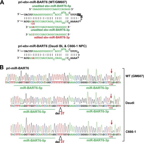
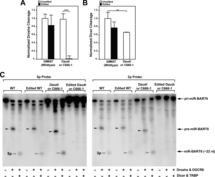
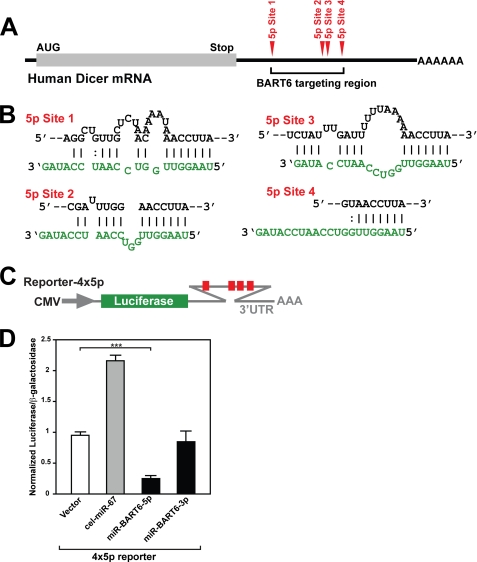
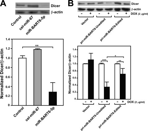
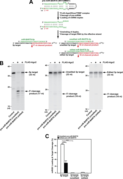
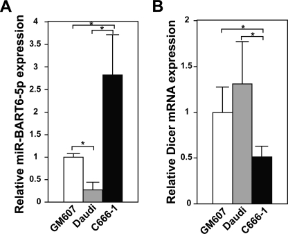
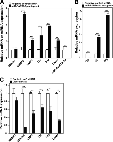
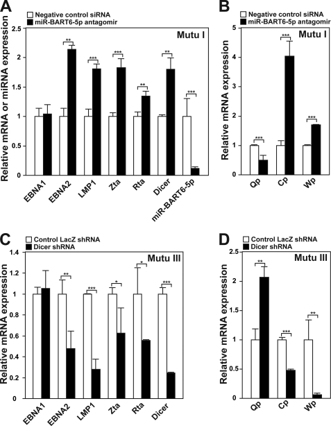
Certain primary transcripts of miRNA (pri-microRNAs) undergo RNA editing that converts adenosine to inosine. The Epstein-Barr virus (EBV) genome encodes multiple microRNA genes of its own. Here we report that primary transcripts of ebv-miR-BART6 (pri-miR-BART6) are edited in latently EBV-infected cells. Editing of wild-type pri-miR-BART6 RNAs dramatically reduced loading of miR-BART6-5p RNAs onto the microRNA-induced silencing complex. Editing of a mutation-containing pri-miR-BART6 found in Daudi Burkitt lymphoma and nasopharyngeal carcinoma C666-1 cell lines suppressed processing of miR-BART6 RNAs. Most importantly, miR-BART6-5p RNAs silence Dicer through multiple target sites located in the 3'-UTR of Dicer mRNA. The significance of miR-BART6 was further investigated in cells in various stages of latency. We found that miR-BART6-5p RNAs suppress the EBNA2 viral oncogene required for transition from immunologically less responsive type I and type II latency to the more immunoreactive type III latency as well as Zta and Rta viral proteins essential for lytic replication, revealing the regulatory function of miR-BART6 in EBV infection and latency. Mutation and A-to-I editing appear to be adaptive mechanisms that antagonize miR-BART6 activities.


Figures

FIGURE 1.
FIGURE 1.
A-to-I RNA editing of pri-miR-BART6 RNAs. A, shown are hairpin structures of pri-miR-BART6. Two different hairpin structures of pri-miR-BART6 (partial), the wild-type from GM607 cells and a mutant found in Daudi Burkitt and C666-1 cells, are shown. The editing site adenosine (+20 site), highlighted in red, is indicated by a number with the 5′ end of the mature miR-BART6–3p sequence counted as +1. The regions to be processed into the mature miRNAs (5p sense and 3p antisense strands) are highlighted in green. Mature miR-BART6-5p and both unedited and edited -3p RNAs are also shown. Three deleted U nucleotides are indicated in black boxes within the wild-type hairpin structure. B, DNA sequencing chromatograms of RT-PCR products derived from GM607, Daudi, and C666-1 pri-miR-BART6 RNAs are shown. The RNA editing site (+20) is detected as an A-to-G change in the cDNA sequencing chromatogram as indicated by red arrows. Three T nucleotides, deleted in pri-miR-BART6 from Daudi and C666-1 cells, are indicated. Editing frequency was estimated as a percentage estimated from the ratio of G peak over the sum of G and A peaks of the sequencing chromatogram. Two separate measurements were done, and identical results were obtained.
FIGURE 2.
FIGURE 2.
In vitro processing of pri-miR-BART6 RNAs by miRNA processor complexes. A, effect of editing on Drosha cleavage of wild-type and mutant pri-miR-BART6 RNAs was tested with uniformly 32P-labeled pri-miR-BART6 RNAs. The mutant pri-miR-BART6 sequences of Daudi and C666-1 are identical. Thus, it is indicated as Daudi or C666-1. Unedited or edited pri-miR-BART6 RNAs (i.e. containing an A-to-G substitution at the +20 site) was subjected to the Drosha cleavage reaction using Drosha-DGCR8 complex. B, effect of editing on Dicer cleavage is shown. The Drosha-DGCR8 reaction products were subjected to the Dicer cleavage reaction using the Dicer-TRBP complex. A and B, three independent assays were done. Differences analyzed by Mann-Whitney U test: **, p < 0.005; ***, p < 0.001. Error bars, S.E. (n = 3). C, Northern blotting analysis of in vitro processed miR-BART6 RNAs is shown. Nonradioactive pri-miR-BART6 RNAs processed in vitro by Drosha-DGCR8 and/or Dicer-TRBP complexes were analyzed by Northern blotting using a 32P-labeled 5p- or 3p-strand specific oligo probe. Representative results for unedited and edited pri-miR-BART6 RNAs of wild-type (GM607) and mutant (Daudi) are shown.
FIGURE 3.
FIGURE 3.
Target sites for miR-BART6-5p RNAs identified in the 3′-UTR of human Dicer mRNA. A, the locations of four miR-BART6-5p target sites located within the 3′-UTR of human Dicer mRNA are schematically presented. B, RNA duplex formation between the Dicer 3′-UTR target sites and miR-BART6-5p RNAs are diagrammed. C, shown is a diagram of the luciferase reporter plasmid containing the four 5p strand target sites. D, relative luciferase activities in HeLa cells cotransfected with the reporter vector containing 4 × 5p sites are shown. Two controls, the vector-only transfection, and cotransfection with the unrelated sequence C. elegans miR-67 were conducted. Expression levels of the luciferase reporter gene were normalized by expression levels of a cotransfected β-galactosidase reporter gene. Three independent assays were conducted. The luciferase activities were compared statistically by Mann-Whitney U tests. Significant differences between vector only and miR-BART6-5p or -3p cotransfected experiments are indicated by asterisks; ***, p < 0.001. Error bars, S.E. (n = 3).
FIGURE 4.
FIGURE 4.
Repression of Dicer by miR-BART6-5p RNAs. A, Western blot analysis of Dicer expression levels in HeLa cells transfected with miR-BART6-5p RNAs is shown. Two control experiments were conducted; HeLa cells without transfection or transfected with a sequence-unrelated C. elegans miR-67. As a normalization control, β-actin levels were also monitored. A summary graph of normalized Dicer expression levels is also presented. B, shown is a Western blot analysis of Dicer expression in HEK293T cells infected with inducible lentivirus vectors for expression of unedited or edited (A-to-G substitution at the +20 site) pri-miR-BART6 RNAs. Expression of pri-miR-BART6 RNAs was induced with 2 μg/ml doxycycline (DOX). In the presence of doxycycline, the control vector directs the expression of non-silencing verified negative siRNAs (Open Biosystems). A summary graph of normalized Dicer expression levels is also shown. A and B, significant differences were analyzed by Mann-Whitney U tests: *, p < 0.05; **, p < 0.005; ***, p < 0.001. Error bars, S.E. (n = 3).
FIGURE 5.
FIGURE 5.
Assembly of functional miRISCs with FLAG-Ago2 and pre-miR-BART6 RNAs. A, a miRISC loading assay of pre-miR-BART6 is shown. Cleavage of the cognate target for miR-BART6-5p or -3p RNAs is schematically shown. The target RNA was 5′ 32P-labeled. B, cleavage of the cognate target product (17 nucleotides) guided by miR-BART6-5p was substantially more efficient with unedited pre-miR-BART6 RNAs than with edited pre-miR-BART6 RNAs (left panel). Cleavage, although very inefficient, of both unedited and edited 3p target was detected only with unedited pre-miR-BART6 (middle and right panels). C, quantitative summary of miRISC loading experiments is presented. The cleavage efficiency was estimated by the ratio of the radioactivity of the correctly cleaved band over that of the uncleaved control band. Significant differences were analyzed by Mann-Whitney U tests: ***, p < 0.001. Error bars, S.E. (n = 3).
FIGURE 6.
FIGURE 6.
Relative expression levels of miR-BART6-5p and Dicer in different cell lines. A, miR-BART6-5p RNA levels were examined by qRT-PCR and normalized to β-actin mRNA level. Three independent assays were done. Significant differences were analyzed by Mann-Whitney U tests. *, p < 0.05. Error bars, S.E. (n = 3). B, Dicer mRNA levels were monitored by qRT-PCR and normalized to β-actin mRNA levels. Three independent assays were performed. Significant differences were analyzed by Mann-Whitney U tests. *, p < 0.05. Error bars, S.E. (n = 3).
FIGURE 7.
FIGURE 7.
Control of viral genes critical for the state of latency and lytic viral replication. A, up-regulation of EBV genes critical for latency and viral replication by the miR-BART6-5p antagomir is shown. Expression of select viral genes including miR-BART6-5p in C666-1 cells transfected with the miR-BART6-5p antagomir or control (sequence unrelated Qiagen AllStars Negative Control siRNA) was examined by qRT-PCR. Three independent assays were done. Significant differences were analyzed by Mann-Whitney U tests. *, p < 0.01; **, p < 0.005; ***, p < 0.001. Error bars, S.E. (n = 3). B, shown are changes induced by the miR-BART6-5p antagomir in the viral promoters Qp, specific for the type I and type II latency, and Cp and Wp, specific for the type III latency. Transcripts initiated from Qp, Cp, and Wp were determined by qRT-PCR and compared with β-actin transcripts. Three independent assays were done. Significant differences were analyzed by Mann-Whitney U tests. **, p < 0.005; ***, p < 0.001. Error bars, S.E. (n = 3). C, repression of EBV genes after Dicer knock-down by shRNA. Expression of viral genes in C666-1 cells transfected with the Dicer targeting shRNA expression plasmid or control vector containing shRNA against LacZ was monitored by qRT-PCR. Three independent assays were conducted. Significant differences were analyzed by the Mann-Whitney U test. *, p < 0.01; ***, p < 0.001. Error bars, S.E. (n = 3).
FIGURE 8.
FIGURE 8.
The effects of miR-BART6-5p and Dicer silencing in B lymphoma cells. A, shown is up-regulation of EBV genes critical for latency III and lytic replication in Mutu I cells transfected with the miR-BART6-5p antagomir or control siRNA. Dicer and miR-BART6-5p levels were also monitored. Three independent qRT-PCR assays were performed. Significant differences were analyzed by Mann-Whitney U tests. **, p < 0.005; ***, p < 0.001. Error bars, S.E. (n = 3). B, changes in viral promoter activities were induced in Mutu I cells by the miR-BART6-5p antagomir. Three independent qRT-PCR assays were done. Significant differences were analyzed by Mann-Whitney U tests. ***, p < 0.001. Error bars, S.E. (n = 3). C, down-regulation of EBV genes critical for latency III and lytic replication was detected in Mutu III cells transfected with the Dicer targeting shRNA expression plasmid or control vector containing shRNA against LacZ. Three independent qRT-PCR assays were done. Significant differences were analyzed by Mann-Whitney U tests. *, p < 0.05; **, <0.005; ***, p < 0.001. Error bars, S.E. (n = 3). D, changes in viral promoter activities were induced in Mutu III cells by Dicer knockdown. Three independent qRT-PCR assays were performed. Significant differences were analyzed by Mann-Whitney U tests. **, p < 0.005; ***, p < 0.001. Error bars, S.E. (n = 3).

Similar articles

Cited by

References

    1. Bartel D. P. (2004) Cell 116, 281–297 - PubMed
    1. Stefani G., Slack F. J. (2008) Nat. Rev. Mol. Cell Biol. 9, 219–230 - PubMed
    1. Esquela-Kerscher A., Slack F. J. (2006) Nat. Rev. Cancer 6, 259–269 - PubMed
    1. Kim V. N., Han J., Siomi M. C. (2009) Nat. Rev. Mol. Cell Biol. 10, 126–139 - PubMed
    1. Winter J., Jung S., Keller S., Gregory R. I., Diederichs S. (2009) Nat. Cell Biol. 11, 228–234 - PubMed

Publication types

MeSH terms

LinkOut - more resources