Review
. 2010 Sep;62(3):525-63.
doi: 10.1124/pr.110.002907.

cGMP-dependent protein kinases and cGMP phosphodiesterases in nitric oxide and cGMP action

Affiliations
Review

cGMP-dependent protein kinases and cGMP phosphodiesterases in nitric oxide and cGMP action

Sharron H Francis et al. Pharmacol Rev. 2010 Sep.

Abstract

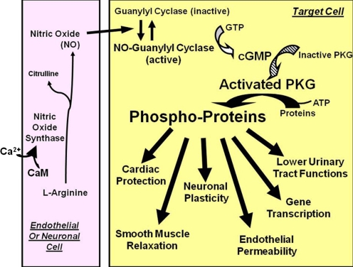
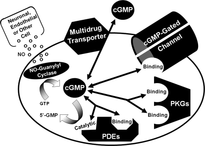
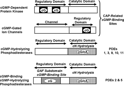
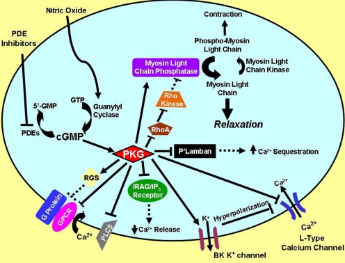
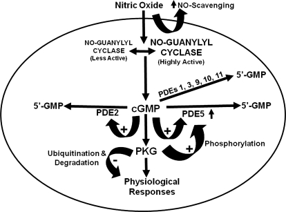
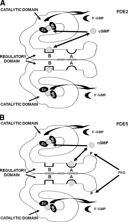
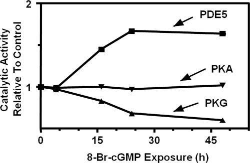
To date, studies suggest that biological signaling by nitric oxide (NO) is primarily mediated by cGMP, which is synthesized by NO-activated guanylyl cyclases and broken down by cyclic nucleotide phosphodiesterases (PDEs). Effects of cGMP occur through three main groups of cellular targets: cGMP-dependent protein kinases (PKGs), cGMP-gated cation channels, and PDEs. cGMP binding activates PKG, which phosphorylates serines and threonines on many cellular proteins, frequently resulting in changes in activity or function, subcellular localization, or regulatory features. The proteins that are so modified by PKG commonly regulate calcium homeostasis, calcium sensitivity of cellular proteins, platelet activation and adhesion, smooth muscle contraction, cardiac function, gene expression, feedback of the NO-signaling pathway, and other processes. Current therapies that have successfully targeted the NO-signaling pathway include nitrovasodilators (nitroglycerin), PDE5 inhibitors [sildenafil (Viagra and Revatio), vardenafil (Levitra), and tadalafil (Cialis and Adcirca)] for treatment of a number of vascular diseases including angina pectoris, erectile dysfunction, and pulmonary hypertension; the PDE3 inhibitors [cilostazol (Pletal) and milrinone (Primacor)] are used for treatment of intermittent claudication and acute heart failure, respectively. Potential for use of these medications in the treatment of other maladies continues to emerge.


Figures

Fig. 1.
Fig. 1.
Nitric oxide signaling through cGMP. NO is synthesized from l-arginine by NO synthases located in neuronal, endothelial, or other cells. Calcium that enters the cell complexes with calmodulin and activates the synthases. The NO produced then diffuses through the intercellular space and traverses the cell membrane of a nearby target cell. Therein, NO binds to and activates NO-guanylyl cyclase, which increases synthesis of cGMP from GTP and results in activation of PKG phosphotransferase activity. These processes initiate a cascade of reactions that are amplified at each step as shown by increasingly larger arrows and text. CaM, calmodulin. [Adapted from Francis SH and Corbin JD (2005) Phosphodiesterase-5 inhibition: the molecular biology of erectile function and dysfunction. Urol Clin North Am 32:419–429. Copyright © 2005 Elsevier Ltd. Used with permission.]
Fig. 2.
Fig. 2.
Cellular targets of cGMP. Multiple intracellular proteins can interact with cGMP, including the cGMP-gated cation channel, PKG, allosteric sites and catalytic sites on certain PDEs, and certain multidrug transporter proteins. The level of cellular cGMP is determined largely by the balance between its synthesis by guanylyl cyclase and breakdown by PDEs, although the transporters may play a role in some cells. The different shapes of the pockets on the respective proteins indicate that the catalytic sites of PDEs (shown as half-diamonds), the allosteric sites of PDEs (shown as half-octagons) and the allosteric sites on PKG and cation channels (shown as half-circles) are structurally and evolutionarily unrelated. The affinity of the multidrug transporter for cGMP is low, and its role in exporting cGMP from the cell is minor compared with the action of PDEs.
Fig. 3.
Fig. 3.
Comparison of structures of major intracellular cGMP receptors. Images demonstrate that PKGs have two allosteric cGMP-binding sites compared with one cGMP-binding site on the subunits of the cGMP-gated channels; the sites on the PKG and the channels belong to the CAP family of cN-binding sites. cGMP-hydrolyzing PDEs contain a catalytic site at which cGMP is converted to 5′-GMP, and some of these PDEs contain an allosteric cGMP-binding site that is located in one of the GAF subdomains found in these proteins. The term GAF is the acronym for a protein domain conserved in cGMP-binding PDEs/Anabaena adenylyl cyclases, and Escherichia coli FhlA.
Fig. 4.
Fig. 4.
Working model of PKGI. PKGI isozymes are homodimers that are dimerized by an extended leucine zipper region (ZZZZZ). Unique faces of the zipper regions that provide for selective homodimerization of PKGIα and PKGIβ and their selective interaction with specific cellular proteins are indicated by the combined asterisks and plus symbols (* + * +). Multiple sites of autophosphorylation are indicated by encircled P and multiple autoinhibitory contacts are indicated by dashed lines (- - - - -). The cGMP-binding sites are homologous, but the more amino-terminal site has higher affinity for cGMP, indicated by the heavy dark semicircle.
Fig. 5.
Fig. 5.
Role of PKGI in modulating intracellular calcium level to promote smooth muscle relaxation. cGMP activation of PKGI in response to nitric oxide elicits multiple phosphorylations of cellular proteins that result in lowering of cellular calcium either through decreased mobilization from intracellular stores or decreased entry from the extracellular space. Solid arrows leading from PKG to a protein indicate that the PKGI-mediated phosphorylation of that protein stimulates its biological activity; for example PKG phosphorylation of RGS proteins activates these proteins, leading to their effect to impair coupling between G proteins and G protein-coupled receptors (GPCRs). An inhibitory bar indicates that the PKGI-mediated phosphorylation impairs the biological function of that protein; for example, PKGI phosphorylation of phospholamban decreases its inhibitory effect on the sarcoplasmic reticulum calcium/ATPase, thereby allowing increased calcium sequestration. PLCβ, phospholipase Cβ. [Adapted from Francis SH and Corbin JD (2005) Phosphodiesterase-5 inhibition: the molecular biology of erectile function and dysfunction. Urol Clin North Am 32:419–429. Copyright © 2005 Elsevier Ltd. Used with permission.]
Fig. 6.
Fig. 6.
Negative feedback mechanisms that blunt or terminate NO and cGMP signaling. cGMP signaling is blunted and/or terminated by multiple points of action that serve to decrease cGMP level by lowering NO-GC activity, activating cGMP-hydrolyzing PDEs, or decreasing levels of the proteins involved in the signaling. PDEs 1, 2, 3, 5, 9, 10, and 11 hydrolyze cGMP and are widespread throughout mammalian tissues. The activities of PDE2 and PDE5 can be further accelerated by allosteric cGMP binding and by phosphorylation for PDE5. [Adapted in part from Francis SH, Corbin JD, and Bischoff E (2009) Cyclic GMP-hydrolyzing phosphodiesterases. Handb Exp Pharmacol 191:367–408. Copyright © 2009 Springer Science+Business Media. Used with permission.]
Fig. 7.
Fig. 7.
Working model of PDE2 and PDE5. A, PDE2 hydrolyzes cGMP and cAMP, and binding of cGMP to GAF-B stimulates cGMP breakdown at the catalytic site, resulting in a negative feedback action of cGMP level. Zn2+ and another divalent metal ion (perhaps Mg2+ or Mn2+) comprise a binuclear metal binding site where a hydroxyl ion from water is polarized for breaking the cyclic phosphate ring. B, PDE5 has overall structure similar to that of PDE2. PKGI phosphorylates Ser-102 located near the amino terminus; both phosphorylation and allosteric cGMP binding, which occurs at GAF-A, increase cGMP breakdown at subsaturating cGMP level. [Adapted from Francis SH and Corbin JD (2005) Phosphodiesterase-5 inhibition: the molecular biology of erectile function and dysfunction. Urol Clin North Am 32:419–429. Copyright © 2005 Elsevier Ltd. Used with permission.]
Fig. 8.
Fig. 8.
Desensitization of cGMP accumulation in response to NO. Strips of aorta were preincubated in the absence [Control (No Pretreatment)] or presence of a NO donor for 10 min; tissue was washed to remove the NO donor. After either 30 or 60 min, the tissue was rechallenged with high levels of the NO donor. Tissues were harvested at indicated times and analyzed for cGMP content. [Adapted from Mullershausen F, Lange A, Mergia E, Friebe A, and Koesling D (2006) Desensitization of NO/cGMP signaling in smooth muscle: blood vessels versus airways. Mol Pharmacol 69:1969–1974. Copyright © 2006 American Society for Pharmacology and Experimental Therapeutics.]
Fig. 9.
Fig. 9.
Long-term regulation of PDE5 and PKGI in response to persistent elevation of cGMP. Cultured rat vascular smooth muscle cells were exposed to 0.1 mM 8-Br-cGMP for various times. PDE5 catalytic activity was measured using 0.3 μM cGMP as substrate ± 0.1 μM sildenafil as described previously (Corbin et al., 2005). PKG and PKA catalytic activities were determined as described previously (Jiang et al., 1992). cAMP-PDE activity [measured at either 0.3 or 30 μM cAMP as described previously (Corbin et al., 2005)] and activity of PDE 1, 3, or 4, determined on the basis of effects of family-selective PDE inhibitors, was unchanged. Prolonged exposure to 0.1 mM 8-Br-cAMP had no effect. Data represent the mean ± S.E. (n = 3–7).

References

    1. Agostino PV, Plano SA, Golombek DA. (2007) Sildenafil accelerates reentrainment of circadian rhythms after advancing light schedules. Proc Natl Acad Sci USA 104:9834–9839 - PMC - PubMed
    1. Ahmad F, Cong LN, Stenson Holst L, Wang LM, Rahn Landstrom T, Pierce JH, Quon MJ, Degerman E, Manganiello VC. (2000) Cyclic nucleotide phosphodiesterase 3B is a downstream target of protein kinase B and may be involved in regulation of effects of protein kinase B on thymidine incorporation in FDCP2 cells. J Immunol 164:4678–4688 - PubMed
    1. Aitken A, Bilham T, Cohen P, Aswad D, Greengard P. (1981) A specific substrate from rabbit cerebellum for guanosine-3′:5′- monophosphate-dependent protein kinase. III. Amino acid sequences at the two phosphorylation sites. J Biol Chem 256:3501–3506 - PubMed
    1. Aitken A, Hemmings BA, Hofmann F. (1984) Identification of the residues on cyclic GMP-dependent protein kinase that are autophosphorylated in the presence of cyclic AMP and cyclic GMP. Biochim Biophys Acta 790:219–225 - PubMed
    1. Aizawa T, Wei H, Miano JM, Abe J, Berk BC, Yan C. (2003) Role of phosphodiesterase 3 in NO/cGMP-mediated antiinflammatory effects in vascular smooth muscle cells. Circ Res 93:406–413 - PubMed

Publication types

MeSH terms

LinkOut - more resources