. 2009 Nov-Dec;2(5):297-306.
doi: 10.4161/oxim.2.5.9541.

Tumor necrosis factor-alpha and apoptosis signal-regulating kinase 1 control reactive oxygen species release, mitochondrial autophagy, and c-Jun N-terminal kinase/p38 phosphorylation during necrotizing enterocolitis

Affiliations
Free PMC article

Tumor necrosis factor-alpha and apoptosis signal-regulating kinase 1 control reactive oxygen species release, mitochondrial autophagy, and c-Jun N-terminal kinase/p38 phosphorylation during necrotizing enterocolitis

Naira Baregamian et al. Oxid Med Cell Longev. 2009 Nov-Dec.
Free PMC article

Abstract

Background: Oxidative stress and inflammation may contribute to the disruption of the protective gut barrier through various mechanisms; mitochondrial dysfunction resulting from inflammatory and oxidative injury may potentially be a significant source of apoptosis during necrotizing enterocolitis (NEC). Tumor necrosis factor (TNF)-alpha is thought to generate reactive oxygen species (ROS) and activate the apoptosis signal-regulating kinase 1 (ASK1)-c-Jun N-terminal kinase (JNK)/p38 pathway. Hence, the focus of our study was to examine the effects of TNF-alpha/ROS on mitochondrial function, ASK1-JNK/p38 cascade activation in intestinal epithelial cells during NEC.

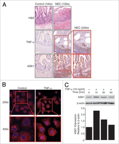
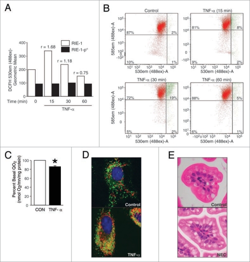
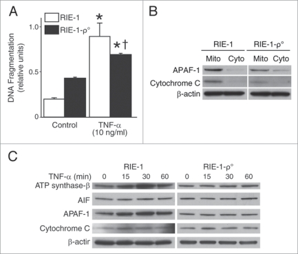
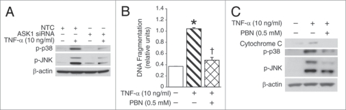
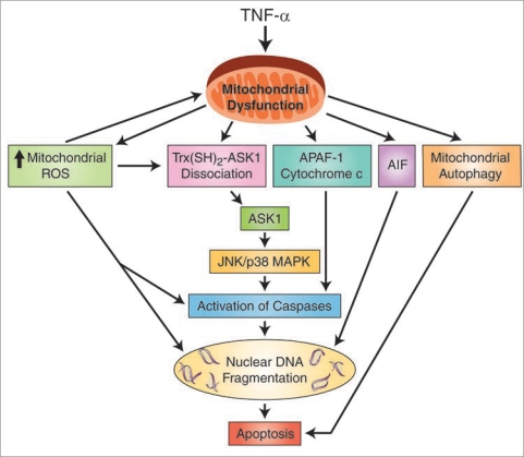
Results: We found (a) abundant tissue TNF-alpha and ASK1 expression throughout all layers of the intestine in neonates with NEC, suggesting that TNF-alpha/ASK1 may be a potential source (indicators) of intestinal injury in neonates with NEC; (b) TNF-alpha-induced rapid and transient activation of JNK/p38 apoptotic signaling in all cell lines suggests that this may be an important molecular characteristic of NEC; (c) TNF-alpha-induced rapid and transient ROS production in RIE-1 cells indicates that mitochondria are the predominant source of ROS, demonstrated by significantly attenuated response in mitochondrial DNA-depleted (RIE-1-rho) intestinal epithelial cells; (d) further studies with mitochondria-targeted antioxidant PBN supported our hypothesis that effective mitochondrial ROS trapping is protective against TNF-alpha/ROS-induced intestinal epithelial cell injury; (e) TNF-alpha induces significant mitochondrial dysfunction in intestinal epithelial cells, resulting in increased production of mtROS, drop in mitochondrial membrane potential (MMP) and decreased oxygen consumption; (f) although the significance of mitochondrial autophagy in NEC has not been unequivocally shown, our studies provide a strong preliminary indication that TNF-alpha/ROS-induced mitochondrial autophagy may play a role in NEC, and this process is a late phenomenon.

Methods: Paraffin-embedded intestinal sections from neonates with NEC and non-inflammatory condition of the gastrointestinal tract undergoing bowel resections were analyzed for TNF-alpha and ASK1 expression. Rat (RIE-1) and mitochondrial DNA-depleted (RIE-1-rho) intestinal epithelial cells were used to determine the effects of TNF-alpha on mitochondrial function.

Conclusions: Our findings suggest that TNF-alpha induces significant mitochondrial dysfunction and activation of mitochondrial apoptotic responses, leading to intestinal epithelial cell apoptosis during NEC. Therapies directed against mitochondria/ROS may provide important therapeutic options, as well as ameliorate intestinal epithelial cell apoptosis during NEC.


Figures

Figure 1
Figure 1
Increased TNFα and ASK1 expression during NEC. (A) The intestinal sections from neonates with NEC and non-inflammatory condition (intestinal atresia; control) were assessed for TNFα and ASK1 expression. Representative section demonstrates villous tip blunting and inflammatory cell infiltration in NEC sections (top). Increased levels of TNFα (middle) and ASK1 (bottom) were localized to mucosa, submucosa and muscular layers in NEC (brown staining). (B) RIE-1 cells (1 × 10⁴/well) were treated with TNFα (10 ng/mL) for 15 min, incubated with anti-ASK1 antibody, AlexaFluor® 647-labeled goat anti-rabbit IgG (pseudo purple) and Hoechst 33342 nuclear stain (blue), and were visualized using confocal microscopy. TNFα treatment resulted in rapid increase in intracellular ASK1 expression in RIE-1 cells. (C) TNFα treatment also increased ASK1 expression in RIE-1 cells by western blotting. Equal β-actin levels and densitometry indicate even loading.
Figure 2
Figure 2
TNFα induces mitochondrial functional deregulation in intestinal epithelial cells. (A) RIE-1 and RIE-1-ρ° cells (1 × 10⁶) were treated with TNFα, incubated with DCFH-DA for 15 min for ROS level. In RIE-1 cells, TNFα treatment induced rapid, transient rise in intracellular ROS. The ratio of stimulated vs. baseline ROS levels increased (r > 1) in TNFα-treated RIE-1 cells only. (B) RIE-1 cells were treated as before and incubated with JC-1 dye for 15 min in darkness. The collapse of the electrochemical gradient across the mitochondrial membrane (red fluorescence) was analyzed by a FACScan flow cytometer. Mitochondria with intact MMP are represented as green fluorescence. Initiation of MMP decrease is seen at 15 min, and recovery at 60 min. (C) RIE-1 cells (1 × 10⁷) were transferred to QO2 chamber and treated with TNFα (10 ng/mL). O2 consumption was significantly decreased by ∼20% shortly after TNFα treatment (*p < 0.05 vs. control). (D) RIE-1 cells (1 × 10⁴/well) were treated with TNFα and incubated with MitoTracker (red fluorescence, mitochondria) and LysoTracker (green fluorescence, lysosomes) dyes. Merged confocal images demonstrated mitochondrial autophagy (yellow fluorescence) in damaged RIE-1 cells at 24 h. (E) Cross-section of H&E-stained neonatal mouse intestinal villi demonstrate autophagic vacuolization of intestinal epithelial cells in NEC group.
Figure 3
Figure 3
TNFα activates mitochondrial apoptotic pathways and induces significant intestinal epithelial cell apoptosis. (A) RIE-1 and RIE-1-ρ° cells were treated with TNFα as before. apoptosis was measured by DNA fragmentation ELISA. Data represent triplicate determinations (mean ± SEM; *p < 0.05 vs. control; †p < 0.05 vs. TNFα-treated RIE-1 cells) and experiments were repeated 3 times. TNFα-induced apoptosis was attenuated when compared to RIE-1 cells. (B) RIE-1 and RIE-1-ρ° cells (2 × 10⁷) were plated for 24 h, mitochondria isolated for protein analysis of mitochondrial apoptotic markers, cytochrome c and APAF-1, by western blotting. Mitochondrial DNA-damaged RIe-1-ρ° cells showed attenuated mitochondrial protein levels. (C) TNFα treatment resulted in increased expression of mitochondrial apoptotic markers (AIF, APAF-1, cytochrome c and ATP synthase-β) in RIE-1 cells by western blotting. Mitochondrial DNA-damaged RIe-1-ρ° cells treated with TNFα showed unchanged basal levels of all apoptotic markers except for increased expression of cytochrome c at 15 min.
Figure 4
Figure 4
ROS trapping and ASK1 silencing during TNFα-induced activation of apoptotic signaling. (A) ASK1 siRNA- and non-targeted control-transfected RIE-1 cells were treated with TNFα for 15 min and cell lysates were analyzed for phosphorylation of JNK and p38 by western blotting. ASK1 silencing resulted in significant reduction in the levels of JNK and p38 activation during TNFα treatment. Equal β-actin levels indicate even loading. (B) RIE-1 cells (2 × 10⁴/well) were pretreated with PBN for 2 h prior to TNFα treatment. PBN pretreatment dramatically reduced TNFα-induced RIe-1 cell death (*p < 0.05 vs. control; †p < 0.05 vs. TNFα alone). (C) TNFα-induced activation of JNK/p38 pathway and cytochrome c expression was signifi- cantly decreased with PBN pretreatment in RIe-1 cells by western blotting.
Figure 5
Figure 5
A proposed mechanism of TNFα-induced mitochondrial dysfunction and activation of apoptotic signaling pathways in the intestinal epithelial cells during NEC. Apoptosis-inducing factor (AIF); apoptotic protease activating factor 1 (APAF-1); apoptosis signal-regulating kinase 1 (ASK1); c-Jun N-terminal kinase (JNK)/p38 pathway; mitogen-activated protein kinase (MAPK); reactive oxygen species (ROS); thioredoxin-apoptosis signal-regulating kinase 1 (Trx(SH)2-ASK1) complex; tumor necrosis factor-α (TNFα).

References

    1. Clark DA, Fornabaio DM, McNeill H, Mullane KM, Caravella SJ, Miller MJ. Contribution of oxygen-derived free radicals to experimental necrotizing enterocolitis. Am J Pathol. 1988;130:537–542. - PMC - PubMed
    1. Kelly N, Friend K, Boyle P, Zhang XR, Wong C, Hackam DJ, et al. The role of the glutathione antioxidant system in gut barrier failure in a rodent model of experimental necrotizing enterocolitis. Surgery. 2004;136:557–566. - PubMed
    1. Martin CR, Walker WA. Intestinal immune defences and the inflammatory response in necrotising enterocolitis. Semin Fetal Neonatal Med. 2006;11:369–377. - PubMed
    1. Markel TA, Crisostomo PR, Wairiuko GM, Pitcher J, Tsai BM, Meldrum DR. Cytokines in necrotizing enterocolitis. Shock. 2006;25:329–337. - PubMed
    1. MacDonald TT, Hutchings P, Choy MY, Murch S, Cooke A. Tumour necrosis factor-alpha and interferon-gamma production measured at the single cell level in normal and inflamed human intestine. Clin Exp Immunol. 1990;81:301–305. - PMC - PubMed

Publication types

MeSH terms

Substances

LinkOut - more resources