Review
. 2010 Mar-Apr;3(2):109-21.
doi: 10.4161/oxim.3.2.11354.

Calcific uremic arteriolopathy: pathophysiology, reactive oxygen species and therapeutic approaches

Affiliations
Free PMC article
Review

Calcific uremic arteriolopathy: pathophysiology, reactive oxygen species and therapeutic approaches

Kurt M Sowers et al. Oxid Med Cell Longev. 2010 Mar-Apr.
Free PMC article

Abstract

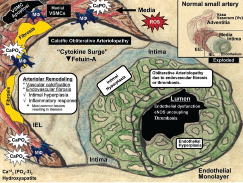
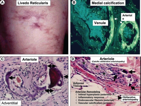
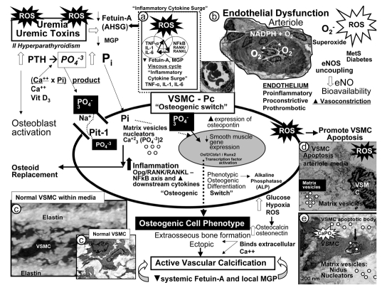
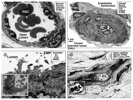
Calcific uremic arteriolopathy (CUA)/calciphylaxis is an important cause of morbidity and mortality in patients with chronic kidney disease requiring renal replacement. Once thought to be rare, it is being increasingly recognized and reported on a global scale. The uremic milieu predisposes to multiple metabolic toxicities including increased levels of reactive oxygen species and inflammation. Increased oxidative stress and inflammation promote this arteriolopathy by adversely affecting endothelial function resulting in a prothrombotic milieu and significant remodeling effects on vascular smooth muscle cells. These arteriolar pathological effects include intimal hyperplasia, inflammation, endovascular fibrosis and vascular smooth muscle cell apoptosis and differentiation into bone forming osteoblast-like cells resulting in medial calcification. Systemic factors promoting this vascular condition include elevated calcium, parathyroid hormone, and hyperphosphatemia with consequent increases in the calcium x phosphate product. The uremic milieu contributes to a marked increased in upstream reactive oxygen species - oxidative stress and subsequent downstream increased inflammation, in part, via activation of the nuclear transcription factor NFkappaB and associated downstream cytokine pathways. Consitutive anti-calcification proteins such as Fetuin-A and matrix GLA proteins and their signaling pathways may be decreased, which further contributes to medial vascular calcification. The resulting clinical entity is painful, debilitating and contributes to the excess morbidity and mortality associated with chronic kidney disease and end stage renal disease. These same histopathologic conditions also occur in patients without uremia and therefore, the term calcific obliterative arteriolopathy could be utilized in these conditions.


Figures

Figure 1
Figure 1
Arteriolar remodeling and vascular calcification in calcific uremic arteriolopathy (CUA)/calciphylaxis. Arteriole model depicted is derived from the pull out model of a normal small artery (upper right insert—boxed in area). This model demonstrates the four most common arteriolar findings observed in histologic sections in CUA/calciphylaxis: Vascular calcification, endovascular fibrosis, intimal hyperplasia, and inflammatory response. Intimal hyperplasia consists of the cellular expansion of the intima including endothelial hyperplasia (green). Excessive reactive oxygen species (ROS) due to uremic toxins may be the driving force promoting this calcific obliterative arteriolopathy due to either endovascular fibrosis or thrombosis. Ca, calcium; EEL, external elastic lamina; eNOS, endothelial derived nitric oxide synthase; IEL, internal elastic lamina; MΦ, macrophage; PO4, phosphate; VSMC, vascular smooth muscle cell.
Figure 2
Figure 2
Early skin changes and histologic findings in calcific uremic arteriolopathy/calciphylaxis. (A) depicts the dermal changes of livedo reticularis (left anterior leg) prior to the initiation of hemodialysis. This image along with painful-palpable subcutaneous masses and plaques represent early skin changes associated with CUA/calciphylaxis. (B) is an inverted colorized hematoxylin and eosin (H&E) stained image, which demonstrates medial calcification (arrows) in an arteriole and adjacent venule. This image is from biopsy of a breast mass one year prior to the development of CUA/calciphylaxis depicted in Figure 3. (C) portrays an outer adventitial location of vascular calcification (arrows) with H&E staining. (D) depicts arteriolar remodeling including intimal hyperplasia, endovascular fibrosis (asterisks) and vascular calcification (arrows) resulting in calcific obliterative arteriolopathy with endothelial fibrosis and arteriolar obliteration. H & E stain.
Figure 3
Figure 3
Intravenous sodium thiosulfate (STS) induced wound healing. Images of CUA eschar (A), clean granulating bed following two weeks of STS (B), healing phase (C) advancing to complete healing 3 months later in a 58 year old female treated with STS (D). Note the proximity of the skin ulceration to the patient's ileostomy and although this ulcer was small, it was highly vulnerable to infection and subsequent sepsis due to proximity to ileostomy. The large subcutaneous palpable nodule (C) was outlined demonstrating its relation to the skin ulceration (∼7 × 14 cm) and gradually regressed after 4 months of STS treatment.
Figure 4
Figure 4
Potential mechanisms involving uremic toxins and reactive oxygen species (ROS) in vascular calcification. Uremic toxins: Increased parathyroid hormone (PTH), phosphorus (Pi) and phosphate (PO4−3), calcium, calcium × phosphorus product, vitamin D3, and ROS significantly contribute to vascular smooth muscle cell (VSMC) and/or pericyte (Pc) differentiation into an osteoblast-like phenotype. Phosphate absorption into these cells is facilitated by the sodium phosphate cotransporter (Pit-1) resulting in an osteogenic switch due to activation of transcription factors: osteoblast-specific cis-acting element (Osf2)—core binding factor alpha1 (Cbfa-1/Runx2). Osteocalcin, osteonectin, bone morphogenic protein-2alpha and alkaline phosphatase (ALP) are inducers of calcification. In contrast, the systemic and local inhibitors of calcification fetuin-A—alpha2-Heremans-Schmid glycoprotein (AHSG) and matrix Gla protein (MGP) are decreased in uremia and calciphylaxis. Further, ROS and inflammatory cytokine surges may contribute to decreased hepatic synthesis of fetuin-A (insert a). Uremic toxins—ROS promote uncoupling of endothelial nitric oxide synthase (eNOS) enzyme via the oxidation of the requisite tetrahydrobiopterin (BH4) cofactor and results in the endothelium becoming a net producer of superoxide—ROS (insert b). Additionally, decreased bioavailable eNO due to eNOS enzyme uncoupling promotes a proinflammatory, proconstrictive, prothrombotic vascular endothelium. ROS are also capable of promoting VSMC apoptosis in the arterial vascular wall (AVW) and when this occurs the matrix vesicles and apoptotic bodies serve as nucleating sites for further calcium deposition in the extracellular matrix of the arteriole media (inserts b–e) (Fig. 1).
Figure 5
Figure 5
Uncoupling of the eNOS enzyme results in the endothelium becoming a net producer of superoxide. This cartoon depicts many of the significant metabolic events leading to endothelial nitric oxide synthase (eNOS) enzyme uncoupling in the endothelium. Reactive oxygen species (ROS) and their oxidative effects of the requisite cofactor tetrahydrobiopterin (BH4) result in eNOS uncoupling. Excessive oxidation of BH4 resulting in the generation of BH3 and BH2 will not run the eNOS reaction to completion. Instead the reaction uncouples and shifts to the C terminal reductase domain and oxygen reacts with the nicotine adenine dinucleotide phosphorus reduced (NADPH) oxidase enzyme resulting in the generation of superoxide [O2]. These dynamic metabolic sequences, involving the uncoupling of the eNOS, reaction result in a proinflammatory, proconstrictive and prothrombotic endothelium, which contributes to endothelial dysfunction. Adapted and expanded with permission.
Figure 6
Figure 6
Potential mechanisms of sodium thiosulfate allowing for its antioxidant, vasodilator and chelation properties. This cartoon demonstrates the molecular structure of sodium thiosulfate (STS) and its two readily donated unpaired electrons, which facilitate quenching of unpaired electrons, generation of the antioxidant glutathione (GSH), vasodilator hydrogen sulfide (H2S), and calcium chelation forming the highly soluble calcium thiosulfate. Adapted with permission.
Figure 7
Figure 7
No vascular calcification following four years of intermittent (3 times/week) intravenous sodium thiosulfate. These histopathologic figures depict numerous open arterioles (arrows) (A–C) with no evidence of calcific obliterative arteriolopathy in the subdermal interstitium from biopsy of skin adjacent to previously healed ulceration in Figure 3. In (D), note the specific stain for calcium (alizarin red) is negative. Insert (d) demonstrates normal periarteriolar adventitial collagen (arrows), while insert (d′) depicts the positive control for alizarin red. Concurrently, this same patient as in Figures 2 and 3 did not have any subcutaneous calcifications when evaluated with bone scan (figure not shown).
Figure 8
Figure 8
Microcirculation ultrastructure in calcific uremic arteriolopathy. (A) depicts a normal small arteriole (approximately 25–30 µm diameter) with normal lining endothelial cell(s) (EC), and a single layer of supportive vascular smooth muscle cell(s) (VSMC), also note the open lumen with numerous red blood cells (RBC), bar = 1 µm. (B) (in contrast) demonstrates a closed arteriolar lumen in a small arteriole (approximately 12–15 µm diameter) from a patient's subcutaneous skin ulceration with CUA compatible with endothelial dysfunction, vasoconstriction, and closed arteriolar lumen (CAL), bar = 1 µm. (C) is a higher magnification of the boxed in region of the endothelium in (B) and may portray an activated endothelium demonstrating multiple cytoplasmic projections containing numerous vesicles, bar = 0.2 µm. Additionally, note the free particles in the lumen, which may represent endothelial microparticles (EMP) from the activated endothelium. Insert (c) displays an arteriole with endothelial denudation (arrows) and abnormal ballooning of ECs with vacuole formation from same patient, bar = 2 µm. (D) depicts an open capillary lumen (CL) in the subcutaneous tissue of skin biopsy adjacent to previous skin ulceration due to CUA (four years earlier, Fig. 3) treated with sodium thiosulfate (STS) for 4 years. Also note the normal appearing pericytes (Pc) and multiple pericyte processes (PcP), which are restored and known to be very sensitive to oxidative stress. Insert (d) portrays a normal open arteriole suggesting that STS may promote both capillary and arteriolar vasodilation.

Similar articles

Cited by

References

    1. Coates T, Kirkland GS, Dymock RB, Murphy BF, Brealey JK, Mathew TH, et al. Cutaneous necrosis from calcific uremic arteriolopathy. Am J Kidney Dis. 1998;32:384–391. - PubMed
    1. Pollock B, Cunliffe W, Merchant W. Calciphylaxis in the absence of renal failure. Clin Exp Dermatol. 2000;25:389. - PubMed
    1. Goyal S, Huhn K, Provost T. Calciphylaxis in a patient without renal failure or elevated parathyroid hormone: the possible aetiological role of chemotherapy. Br J Dermatol. 2000;143:1087–1090. - PubMed
    1. Nigwekar SU, Wolf M, Sterns RH, Hix JK. Calciphylaxis from nonuremic causes: a systematic review. Clin J Am Soc Nephrol. 2008;3:1139–1143. - PMC - PubMed
    1. Don BR, Chin AL. A strategy for the treatment of calcific uremic arteriolopathy (calciphylaxis) employing a combination of therapies. Clin Nephrol. 2003;59:463–470. - PubMed

Publication types

Substances

LinkOut - more resources