. 2010 May 12;11(3):3274.
doi: 10.1120/jacmp.v11i3.3274.

A comprehensive study on decreasing the kilovoltage cone-beam CT dose by reducing the projection number

Affiliations

A comprehensive study on decreasing the kilovoltage cone-beam CT dose by reducing the projection number

Bo Lu et al. J Appl Clin Med Phys. .

Abstract

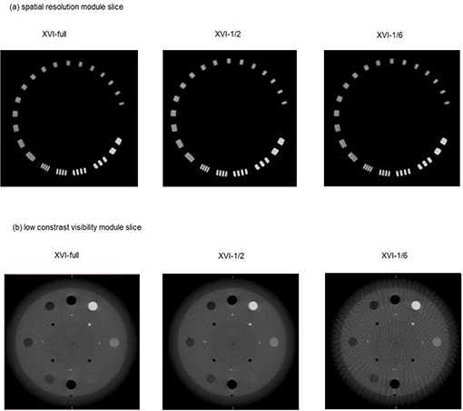
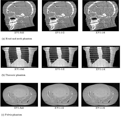
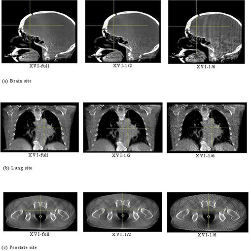
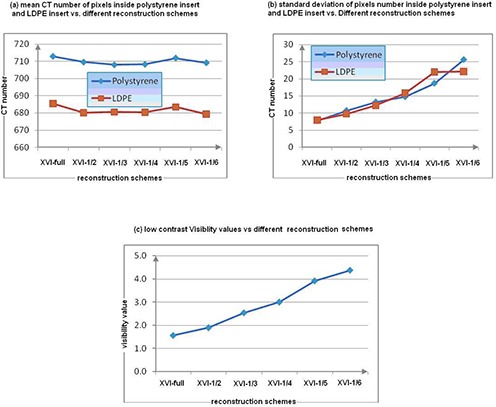
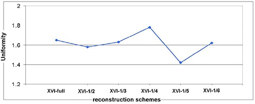
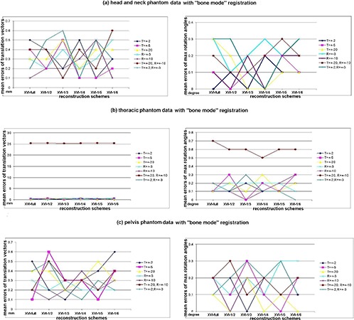
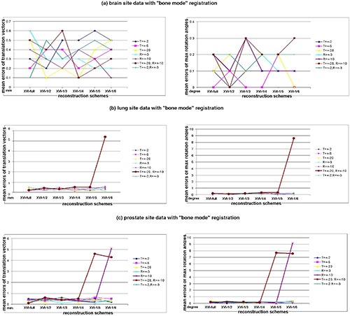
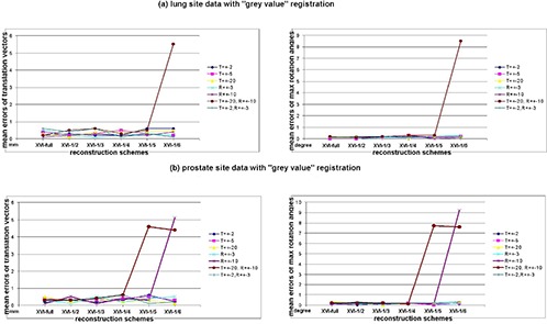
The objective of this study was to evaluate the effect of kilovoltage cone-beam computed tomography (CBCT) on registration accuracy and image qualities with a reduced number of planar projections used in volumetric imaging reconstruction. The ultimate goal is to evaluate the possibility of reducing the patient dose while maintaining registration accuracy under different projection-number schemes for various clinical sites. An Elekta Synergy Linear accelerator with an onboard CBCT system was used in this study. The quality of the Elekta XVI cone-beam three-dimensional volumetric images reconstructed with a decreasing number of projections was quantitatively evaluated by a Catphan phantom. Subsequently, we tested the registration accuracy of imaging data sets on three rigid anthropomorphic phantoms and three real patient sites under the reduced projection-number (as low as 1/6th) reconstruction of CBCT data with different rectilinear shifts and rota-tions. CBCT scan results of the Catphan phantom indicated the CBCT images got noisier when the number of projections was reduced, but their spatial resolution and uniformity were hardly affected. The maximum registration errors under the small amount transformation of the reference CT images were found to be within 0.7 mm translation and 0.3 masculine rotation. However, when the projection number was lower than one-fourth of the full set with a large amount of transformation of reference CT images, the registration could easily be trapped into local minima solutions for a nonrigid anatomy. We concluded, by using projection-number reduction strategy under conscientious care, imaging-guided localization procedure could achieve a lower patient dose without losing the registration accuracy for various clinical sites and situations. A faster scanning time is the main advantage compared to the mA decrease-based, dose-reduction method.


Figures

Figure 1
Figure 1
A spatial‐resolution module slice (a) of a Catphan phantom image reconstructed with XVI‐full, XVI‐1/2, and XVI‐1/6 schemes; a low‐contrast visibility module slice (b) of a Catphan phantom image reconstructed with XVI‐full, XVI‐1/2, and XVI‐1/6 schemes.
Figure 2
Figure 2
Head‐and‐neck phantom (a) with images of a RANDO phantom including XVI‐full, XVI‐1/2, and XVI‐1/6 reconstruction image slices in the sagittal view; thoracic phantom (b) with images of a RANDO phantom including XVI‐full, XVI‐1/2, and XVI‐1/6 reconstruction image slices in the coronal view; pelvis phantom (c) with images of the pelvis area of a RANDO phantom including XVI‐full, XVI‐1/2, and XVI‐1/6 reconstruction image slices in the axial view.
Figure 3
Figure 3
Brain site (a): the patient images of the brain site including XVI‐full, XVI‐1/2, and XVI‐1/6 reconstruction image slices in the sagittal view; lung site (b): patient images of the lung site including XVI‐full, XVI‐1/2, and XVI‐1/6 reconstruction image slices in the coronal view; prostate site (c): patient images of the prostate site including XVI‐full, XVI‐1/2, and XVI‐1/6 reconstruction image slices in the axial view.
Figure 4
Figure 4
Results of a low‐contrast visibility check with different reconstruction schemes.
Figure 5
Figure 5
Results of a uniformity check with different reconstruction schemes.
Figure 6
Figure 6
The scatter charts of mean registration errors of the translation vector and mean registration errors of maximum rotation angles of all directions versus reconstruction schemes with different translation and rotation scales for phantoms with “bone mode” registration algorithm: (a) head‐and‐neck phantom data with the “bone mode” registration; (b) thoracic phantom data with the “bone mode” registration; (c) pelvis phantom data with the “bone mode” registration. Note: all mean registration error values of the translation vector were rounded to one‐tenth of a millimeter; all mean registration error values of maximum rotation angles were rounded to one‐tenth of a degree.
Figure 7
Figure 7
The scatter charts of mean registration errors of the translation vector and mean registration errors of maximum rotation angles of all directions versus reconstruction schemes with different translation and rotation scales for all patient sites with “bone mode” registration algorithm: (a) brain site data with the “bone mode” registration; (b) lung site data with the “bone mode” registration; (c) prostate site data with the “bone mode” registration. Note: all mean registration error values of the translation vector were rounded to one‐tenth of a millimeter; all mean registration error values of maximum rotation angles were rounded to one‐tenth of a degree.
Figure 8
Figure 8
The scatter charts of mean registration errors of the translation vector and mean registration errors of maximum rotation angles of all directions versus reconstruction schemes with different translation and rotation scales for non‐rigid patient sites with “grey value” registration algorithm: (a) lung site data with the “grey value” registration; (b) prostate site data with the “grey value” registration. Note: all mean registration error values of the translation vector were rounded to one‐tenth of a millimeter; all mean registration error values of maximum rotation angles were rounded to one‐tenth of a degree.

Similar articles

Cited by

References

    1. Chauvet B, de Rauglaudre G, Mineur L, Alfonsi M. Reboul F. [Dose‐response relationship in radiotherapy: an evidence?]. Cancer Radiother. 2003;7 Suppl 1:8s–14s. - PubMed
    1. Islam MK, Purdie TG, Norrlinger BD, et al. Patient dose from kilovoltage cone beam computed tomography imaging in radiation therapy. Med Phys. 2006;33(6):1573–82. - PubMed
    1. Pisani L, Lockman D, Jaffray D, Yan D, Martinez A, Wong, J . Setup error in radiotherapy: on‐line correction using electronic kilovoltage and megavoltage radiographs. Int J Radiat Oncol Biol Phys. 2000;47(3):825–39. - PubMed
    1. Jaffray DA, Drake DG, Moreau M, Martinez AA, Wong, JW . A radiographic and tomographic imaging system integrated into a medical linear accelerator for localization of bone and soft‐tissue targets. Int J Radiat Oncol Biol Phys. 1999;45(3):773–89. - PubMed
    1. Jaffray DA, Siewerdsen JH. Cone‐beam computed tomography with a flat‐panel imager: initial performance characterization. Med Phys. 2000;27(6):1311–23. - PubMed

MeSH terms