. 2010 Nov;18(11):1896-906.
doi: 10.1038/mt.2010.169. Epub 2010 Aug 17.

Hyperactive sleeping beauty transposase enables persistent phenotypic correction in mice and a canine model for hemophilia B

Affiliations

Hyperactive sleeping beauty transposase enables persistent phenotypic correction in mice and a canine model for hemophilia B

Martin A Hausl et al. Mol Ther. 2010 Nov.

Abstract

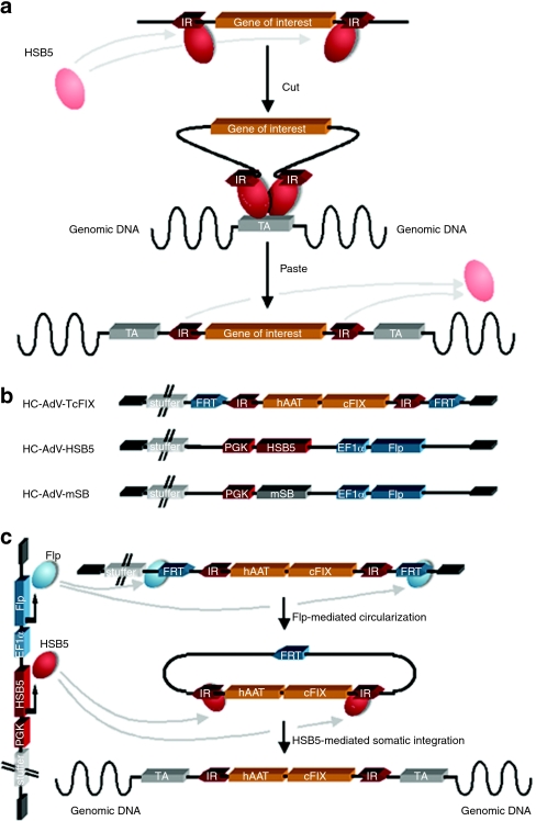
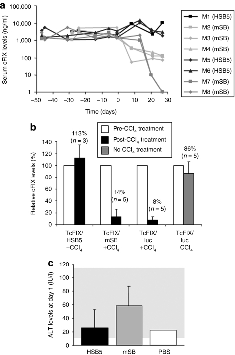
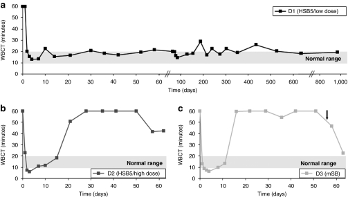
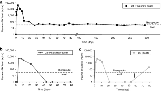
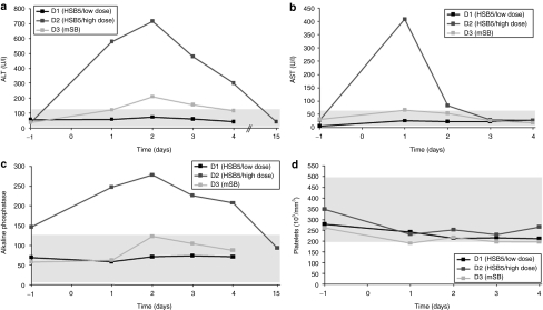
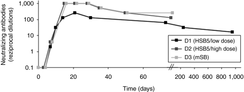
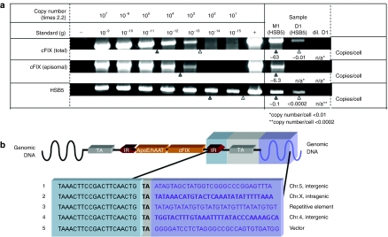
Sleeping Beauty (SB) transposase enables somatic integration of exogenous DNA in mammalian cells, but potency as a gene transfer vector especially in large mammals has been lacking. Herein, we show that hyperactive transposase system delivered by high-capacity adenoviral vectors (HC-AdVs) can result in somatic integration of a canine factor IX (cFIX) expression-cassette in canine liver, facilitating stabilized transgene expression and persistent haemostatic correction of canine hemophilia B with negligible toxicity. We observed stabilized cFIX expression levels during rapid cell cycling in mice and phenotypic correction of the bleeding diathesis in hemophilia B dogs for up to 960 days. In contrast, systemic administration of an inactive transposase system resulted in rapid loss of transgene expression and transient phenotypic correction. Notably, in dogs a higher viral dose of the active SB transposase system resulted into transient phenotypic correction accompanied by transient increase of liver enzymes. Molecular analysis of liver samples revealed SB-mediated integration and provide evidence that transgene expression was derived mainly from integrated vector forms. Demonstrating that a viral vector system can deliver clinically relevant levels of a therapeutic protein in a large animal model of human disease paves a new path toward the possible cure of genetic diseases.


Figures

Figure 1
Figure 1
Mechanism of Sleeping Beauty (SB) transposition and the high-capacity adenoviral vector (HC-AdV) system for delivery. (a) Depicted is a two-component system in which a gene of interest flanked by transposon-derived inverted repeats (IRs) is mobilized by the transposase protein provided in trans. For gene transfer experiments, the transposon is usually excised from a plasmid and integrated into a genomic target site (TA). This mechanism is commonly referred to as “cut-and-paste” mechanism. (b) HC-AdV-TcFIX represents the transposon-donor vector from which the transposon is mobilized. The transposon15 flanked by inverted repeats (IRs) and FRT sites for Flp-mediated excision. It expresses canine coagulation factor IX (cFIX) under control of the liver-specific human α-1-antitrypsin promoter (hAAT) including two liver-specific enhancers (HCR, hepatocyte control region; ApoE: apolipoprotein E). The HC-AdV-HSB5 contains a transgene expression-cassette for the hyperactive Sleeping Beauty (SB) transposase HSB5 under the control of the phosphoglycerate kinase promoter (PGK) and an expression-cassette for the Flp recombinase driven by the elongation factor-1-α promoter (EF1α). The control vector HC-AdV-mSB contains the mutated and inactive version of SB (mSB). All HC-AdVs include 22-kb stuffer DNA derived from human chromosomal DNA to optimize packaging of viral vectors. (c) For somatic integration cells are simultaneously infected with HC-AdV-TcFIX and HC-AdV-HSB5. Expressed Flp-recombinase recognizes FRT sites and mediates circularization and therefore transposon mobilization from the HC-AdV, which is essential for HSB5 functionality. Inverted repeats (IRs) of circularized intermediates are recognized by expressed hyperactive SB protein provided in trans, mediating somatic integration of the transposon into the cellular genome. HC-AdV, high-capacity adenoviral vector; HSB5, hyperactive Sleeping Beauty transposase; mSB, inactive transposase system.
Figure 2
Figure 2
Stability of expression levels in mice treated with adenovirus-transposase hybrid-vectors and acute toxicity related with vectors. C57Bl/6 mice were coinjected with HC-AdV-TcFIX and HC-AdV-HSB5 at a ratio of 3:1 with a total number of 8 × 10¹⁰ viral particles (mouse M1) or 4 × 10¹⁰ viral particles (mice M5 and M6). Mice of the control groups were treated with HC-AdV-mSB instead of HC-AdV-HSB5 (8 × 10¹⁰ viral particles: mice M2–M4; 4 × 10¹⁰ viral particles: mice M7 and M8). Four to seven weeks after vector administration rapid hepatocyte proliferation was induced at day 0 by intraperitonal injection of carbon tetrachloride (CCl4) to analyze stability of transgene expression and somatic integration. (a) Canine FIX expression levels were measured periodically by ELISA. (b) Relative transgene expression levels in mice after delivery of HC-AdV-TcFIX with either HC-AdV-HSB5 (TcFIX/HSB5 + CCl4), HC-AdV-mSB (TcFIX/mSB + CCl4) or the irrelevant vector HC-AdV-luciferase (TcFIX/Luc + CCl4) as a control for persistence of the canine FIX-expressing vector without excision of the transgene by Flp recombination. To analyze persistence of transgene expression without CCl4 treatment (TcFIX/luc −CCl4), another control group solely received the vector HC-AdV-TcFIX and HC-AdV-luciferase. A total of 8 × 10⁸ transducing units were injected at a ratio of 3:1 (transposon TcFIX vector: HSB5, mSB, or Luc vector). Mice were treated with CCl4 and serum levels of cFIX before CCl4 treatment (white bar) were defined as 100% for each group. Black bar, post-CCl4 treatment; gray bar: no CCl4 treatment. Serum cFIX levels were determined 3 weeks after CCl4 treatment for all groups. (c) Alanine transaminase (ALT) levels in serum samples of mice 1 day postinjection were measured to detect acute liver toxicity related with HC-AdV administration. Serum samples of mice injected with Dulbecco's phosphate-buffered saline (DPBS), the solvent for intravenous application of HC-AdVs were used as a negative control. cFIX, canine factor IX; ELISA, enzyme-linked immunosorbent assay; HC-AdV, high-capacity adenoviral vector; HSB5, hyperactive Sleeping Beauty transposase; mSB, inactive transposase system; PBS, phosphate-buffered saline.
Figure 3
Figure 3
Whole blood clotting time (WBCT) as indicator for phenotypic correction in treated hemophilia B dogs. The whole blood clotting time (WBCT) is an indicator for a functional coagulation cascade and therefore for a sufficient replacement of the inactive factor IX in hemophilia B dogs. The WBCT for untreated hemophilia B dogs is longer than 60 minutes. After injection of HC-AdVs the WBCT was measured periodically for (a) D1 (HSB5/low dose), (b) D2 (HSB5/high dose), and (c) D3 (mSB). The black arrow indicates a bleeding episode, which was treated by cFIX administration. cFIX, canine factor IX; HC-AdV, high-capacity adenoviral vector; HSB5, hyperactive Sleeping Beauty transposase; mSB, inactive transposase system.
Figure 4
Figure 4
Expression levels of canine factor IX in treated hemophilia B dogs. The expression level of canine factor IX (cFIX), which is encoded by the transposon, was measured by ELISA. Normal cFIX plasma levels of healthy dogs are between 500 and 12,000 ng/ml. Notably, 50 ng/ml are sufficient for effective coagulation (therapeutic level). Plasma levels of cFIX were measured in hemophilia B dog (a) D1 (HSB5/low dose), (b) D2 (HSB5/high dose), and (c) D3 (mSB). The black arrow indicates the administration of cFIX after a bleeding episode. ELISA, enzyme-linked immunosorbent assay; HSB5, hyperactive Sleeping Beauty transposase; mSB, inactive transposase system.
Figure 5
Figure 5
Acute toxicity profile of treated hemophilia B dogs. Plasma samples of dogs D1 (HSB5/low dose), D2 (HSB5/high dose), and D3 (mSB) were collected at indicated time points and analyzed for markers related with acute liver toxicity. (a) Alanine aminotransferase levels (ALT; normal range 12–118 U/l) were measured as well as (b) aspartate aminotransferase levels (AST; normal range 15–66 U/l), and (c) alkaline phosphatase levels (normal range 5–131 U/l). In addition (d) the number of platelets (normal range 200,000–500,000 per mm3) were quantified for all three dogs. HSB5, hyperactive Sleeping Beauty transposase; mSB, inactive transposase system.
Figure 6
Figure 6
Neutralizing antibodies directed against adenoviral vectors in treated hemophilia B dogs. As an indicator for the adaptive immune response challenged by the HC-AdVs, neutralizing antibodies directed against the vector were measured. Therefore, the highest dilution of canine serum was determined, which is able to inhibit 50% transduction efficiencies of a reporter virus in 293 cells. The neutralizing-antibody titer was defined as the reciprocal of this dilution. HC-AdV, high-capacity adenoviral vector.
Figure 7
Figure 7
Molecular status of vector genomes and detection of transposition events in transduced liver cells. Hyperactive Sleeping Beauty transposase (HSB5)-transduced liver samples of mouse M1 (28 days post-CCl4 treatment) and dog D1 (960 days postinjection) were analyzed regarding episomal HC-AdV genomes and canine factor IX (cFIX) transposon integrated into the host genome. (a) Fate of vector genomes in transduced canine and murine liver was analyzed by PCR. Top, total number of coagulation factor IX transgenes; middle, detection of intact and episomal transposase donor-vector genomes (high-capacity adenoviral vector HC-AdV-TcFIX); bottom, PCR specifically detecting Sleeping Beauty transposase encoding sequences. D1, liver genomic DNA from dog D1 (500 ng); dil. D1, liver genomic DNA from dog D1 (10 ng); M1, liver genomic DNA from mouse M1 (10 ng). Serial DNA standard dilutions ranged from 1 ng (10−9 g; 2.2 × 10⁷ genome copies) to 1 fg (10−15 g; 22 genome copies) were analyzed and spiked with 50-ng genomic DNA derived from 293 cells to simulate canine genomic DNA in samples. Genomic DNA obtained from HeLa cells, infected with the corresponding viral vectors was used as positive control (+) whereas DNA from noninfected HeLa cells served as negative control (–). Vector genome copies are presented as copy numbers per liver cell (copy number/cell). (b) Identification of sites of insertion in canine liver cell genomes after HSB5-mediated transposition. Genomic regions containing the transposon were isolated, sequenced, and blasted against the canine genome database. Five integration events are depicted and the corresponding chromosomal regions are mentioned. HC-AdV, high-capacity adenoviral vector.

Similar articles

Cited by

References

    1. Kay MA, Rothenberg S, Landen CN, Bellinger DA, Leland F, Toman C, et al. In vivo gene therapy of hemophilia B: sustained partial correction in factor IX-deficient dogs. Science. 1993;262:117–119. - PubMed
    1. Brown BD, Cantore A, Annoni A, Sergi LS, Lombardo A, Della Valle P, et al. A microRNA-regulated lentiviral vector mediates stable correction of hemophilia B mice. Blood. 2007;110:4144–4152. - PubMed
    1. Vandendriessche T, Thorrez L, Acosta-Sanchez A, Petrus I, Wang L, Ma L, et al. Efficacy and safety of adeno-associated viral vectors based on serotype 8 and 9 vs. lentiviral vectors for hemophilia B gene therapy. J Thromb Haemost. 2007;5:16–24. - PubMed
    1. Niemeyer GP, Herzog RW, Mount J, Arruda VR, Tillson DM, Hathcock J, et al. Long-term correction of inhibitor-prone hemophilia B dogs treated with liver-directed AAV2-mediated factor IX gene therapy. Blood. 2009;113:797–806. - PMC - PubMed
    1. Manno CS, Pierce GF, Arruda VR, Glader B, Ragni M, Rasko JJ, et al. Successful transduction of liver in hemophilia by AAV-Factor IX and limitations imposed by the host immune response. Nat Med. 2006;12:342–347. - PubMed

Publication types