Review
. 2010 Aug 18;29(16):2677-88.
doi: 10.1038/emboj.2010.175.

Spatial organization of transmembrane receptor signalling

Affiliations
Review

Spatial organization of transmembrane receptor signalling

Ioanna Bethani et al. EMBO J. .

Abstract

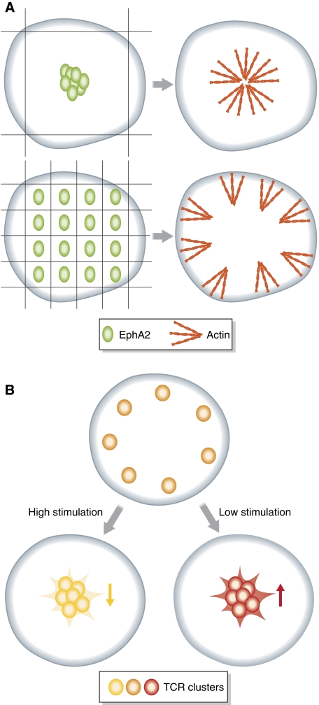
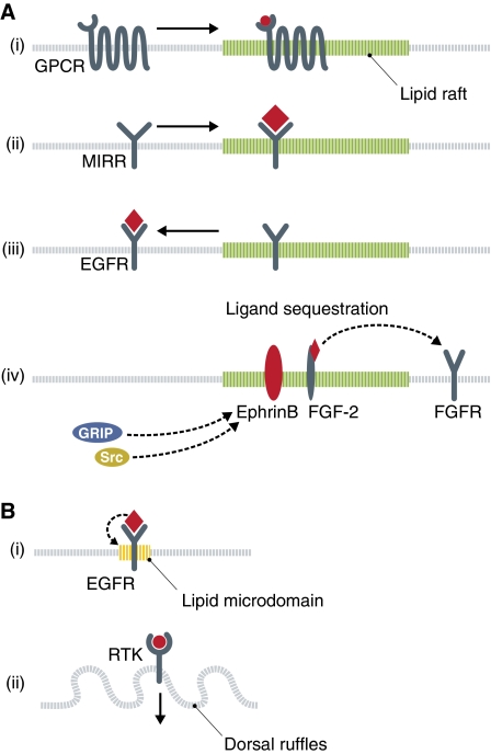
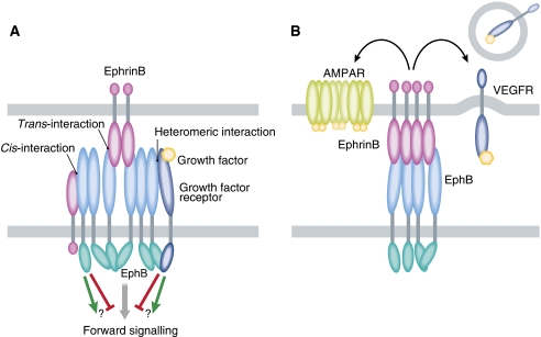
The spatial organization of transmembrane receptors is a critical step in signal transduction and receptor trafficking in cells. Transmembrane receptors engage in lateral homotypic and heterotypic cis-interactions as well as intercellular trans-interactions that result in the formation of signalling foci for the initiation of different signalling networks. Several aspects of ligand-induced receptor clustering and association with signalling proteins are also influenced by the lipid composition of membranes. Thus, lipid microdomains have a function in tuning the activity of many transmembrane receptors by positively or negatively affecting receptor clustering and signal transduction. We review the current knowledge about the functions of clustering of transmembrane receptors and lipid-protein interactions important for the spatial organization of signalling at the membrane.


Conflict of interest statement

The authors declare that they have no conflict of interest.

Figures

Figure 1
Figure 1
Spatial distribution of membrane receptors. (A) The size and organization of EphA2 membrane clusters determines the distribution of downstream effectors. Under conditions of unrestricted membrane transport of EphA2 receptor (green), f-actin (red) accumulates in the periphery of activated ephrinA1–EphA2 clusters. Upon introduction of a spatial mutation that restricts EphA2 organization at the membrane, the distribution of f-actin shifted to the cell periphery (Salaita et al, 2010). (B) Re-distribution of T-cell receptors (TCRs) to the centre of the immunological synapse results in different signalling states depending on the stimulus strength. Upon high stimulation, transport of TCRs (yellow) to the centre of the synapse results in receptor deactivation and attenuation of signalling (pale yellow receptors). In contrast, under low-stimulation, the artificially-forced translocation of TCRs to the centre of the synapse enhances downstream signalling.
Figure 2
Figure 2
Lipid microdomains in receptor organization and signalling. (A) Lipid rafts (green) can serve to recruit ligand (red) stimulated receptors, such as (i) G-protein-coupled receptors (GPCRs) or (ii) multichain immune recognition receptors (MIRRs) and thus focus the downstream signalling complexes. (iii) The receptor tyrosine kinase (RTK) epidermal growth factor receptor (EGFR) is localized to rafts in resting cells and is transported out of the raft upon ligand binding. (iv) A third function for rafts in signal transduction is to sequester receptor ligands. EphrinB is a transmembrane ligand associated with rafts, and serves to recruit cytoplasmic effector molecules to this domain. The FGF-2 ligand is kept in rafts by glypican-1 and away from the non-raft-associated fibroblast growth factor receptor (FGFR). (B) Other microdomains in the membrane can also serve to spatially arrange transmembrane receptors. (i) Upon ligand stimulation, the EGFR induces the formation of a local lipid domain rich in phosphatidic acid (yellow), which also contains an elevated number of EGFRs. (ii) Dorsal ruffles can be induced by stimulation of RTKs. These domains may serve both as signalling platforms and as internalization sites.
Figure 3
Figure 3
Receptor–receptor complexes regulate signalling specificity and receptor trafficking. (A) Eph receptors (Ephs) are clustered on the membrane through trans-interactions with pre-clustered ephrin ligands or through homomeric interactions of their extracellular or intracellular domains. Clustering is necessary for the receptor activation and signalling. The downstream signalling of activated Ephs can be further modulated by interactions with ephrin ligands in cis or by heteromeric associations with other receptor types. (B) EphrinB2 differentially regulates the trafficking of AMPA and VEGF receptors. Serine phosphorylation of ephrinB2 promotes AMPAR stabilization at the cellular membrane of neurons, whereas ephrinB2 positively regulates VEGFR endocytosis through its PDZ-binding domain.

Similar articles

Cited by

References

    1. Abella JV, Parachoniak CA, Sangwan V, Park M (2010) Dorsal ruffle microdomains potentiate Met RTK signalling and downregulation. J Biol Chem (e-pub ahead of print) - PMC - PubMed
    1. Alves ID, Salamon Z, Hruby VJ, Tollin G (2005) Ligand modulation of lateral segregation of a G-protein-coupled receptor into lipid microdomains in sphingomyelin/phosphatidylcholine solid-supported bilayers. Biochemistry 44: 9168–9178 - PMC - PubMed
    1. Ames P, Studdert CA, Reiser RH, Parkinson JS (2002) Collaborative signaling by mixed chemoreceptor teams in Escherichia coli. Proc Natl Acad Sci USA 99: 7060–7065 - PMC - PubMed
    1. Ariotti N, Liang H, Xu Y, Zhang Y, Yonekubo Y, Inder K, Du G, Parton RG, Hancock JF, Plowman SJ (2010) EGFR activation remodels the plasma membrane lipid environment to induce nanocluster formation. Mol Cell Biol 30: 3795–3804 - PMC - PubMed
    1. Arnaout MA, Mahalingam B, Xiong JP (2005) Integrin structure, allostery, and bidirectional signaling. Annu Rev Cell Dev Biol 21: 381–410 - PubMed

Publication types

Substances