Review
. 2010 Aug 18;29(16):2715-23.
doi: 10.1038/emboj.2010.177.

An intimate liaison: spatial organization of the endoplasmic reticulum-mitochondria relationship

Affiliations
Review

An intimate liaison: spatial organization of the endoplasmic reticulum-mitochondria relationship

Olga Martins de Brito et al. EMBO J. .

Abstract

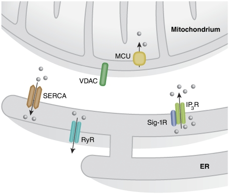
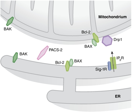
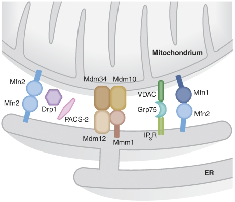
Organelle localization is often crucial to properly modulate cellular functions and signalling cascades. For example, the distribution of organelles in axons is crucial for their function and is dysregulated in several diseases. Similarly, relative positioning of two or more organelles is also important to perform certain specialized processes. Perhaps, the best-known form of interorganellar organization is that between endoplasmic reticulum (ER) and mitochondria. Close communication between these two compartments has been observed for a long time. Recent evidence suggests that this is the basis for a bidirectional communication regulating a number of physiological processes ranging from mitochondrial energy and lipid metabolism to Ca(2+) signalling and cell death. The recent discovery of some of the molecular mediators of the tethering already allowed to extend the function of this paradigmatic spatial organization to previously unexpected functions, and will foster future research to explore it in cellular signalling cascades as well as in disease.


Conflict of interest statement

The authors declare that they have no conflict of interest.

Figures

Figure 1
Figure 1
Local Ca2+ signalling at the ER–mitochondria interface. The proteins involved in Ca2+ signalling between ER and mitochondria are shown. Ca2+ release from the ER occurs mainly through IP3R and RyR enriched at the regions of the ER in close contact with mitochondria. These receptors account for the formation of microdomains of high Ca2+ concentration that are needed to activate the transport of the ion into the mitochondrial matrix through the MCU. Finally, the recently identified Sig-1R is able to modulate the activity of the IP3R and thus Ca2+ transmission from the ER to mitochondria.
Figure 2
Figure 2
Apoptotic signalling at the ER–mitochondria interface. Cross-talk between ER and mitochondria has a major function in the decision whether the cell should live or die. Bcl-2 family members can control apoptosis by controlling indirectly the amount of ER-releasable Ca2+ that can reach mitochondria. Drp1 has a dual function in controlling apoptosis. On one side, recruitment of Drp1 to mitochondria upon sustained Ca2+ release from the ER can protect from cell death by fragmenting the mitochondrial network and impeding the propagation of the fatal Ca2+ wave. On the other hand, mobilization of Drp1 to mitochondria can also trigger mitochondrial cristae remodelling, facilitating cytochrome c mobilization and subsequent apoptosis. Also, the recently identified Sig-1R has a bivalent function in apoptosis. Sig-1R promotes Ca2+ transmission to mitochondria through the IP3R thus maintaining mitochondrial metabolism in conditions of ER Ca2+ depletion. However, excessive Ca2+ transfer to mitochondria risks to expose this organelle to Ca2+ overload and subsequent dysfunction. In this regard, the truncated version of SERCA-1 S1T, expressed upon ER stress, promotes Ca2+ transfer to mitochondria leading to Ca2+ overload.
Figure 3
Figure 3
Tethers between ER and mitochondria. The molecular bridges that regulate the close contacts between ER and mitochondria are shown. PACS-2 and Drp1 indirectly controls the distance between the two organelles by impinging on mitochondrial morphology and distribution. A more direct function in linking ER and mitochondria has been suggested for the complex composed by the IP3R on the ER, the cytosolic chaperone Grp75 and the mitochondrial anion channel VDAC. Further, ERMES, a multimeric complex formed by the mitochondrial proteins Mdm34 and Mdm10 and Mmm1 and Mdm12 on the ER appears to regulate ER–mitochondria tethering in yeast. Importantly, the dynamin-related GTPase Mfn2 on the ER forms homo–heterodimers with Mfn1 or Mfn2 on mitochondria to keep the tight contacts between the two organelles.

Similar articles

Cited by

References

    1. Addya S, Anandatheerthavarada HK, Biswas G, Bhagwat SV, Mullick J, Avadhani NG (1997) Targeting of NH2-terminal-processed microsomal protein to mitochondria: a novel pathway for the biogenesis of hepatic mitochondrial P450MT2. J Cell Biol 139: 589–599 - PMC - PubMed
    1. Amarilio R, Ramachandran S, Sabanay H, Lev S (2005) Differential regulation of endoplasmic reticulum structure through VAP-Nir protein interaction. J Biol Chem 280: 5934–5944 - PubMed
    1. Anandatheerthavarada HK, Biswas G, Mullick J, Sepuri NB, Otvos L, Pain D, Avadhani NG (1999) Dual targeting of cytochrome P4502B1 to endoplasmic reticulum and mitochondria involves a novel signal activation by cyclic AMP-dependent phosphorylation at ser128. EMBO J 18: 5494–5504 - PMC - PubMed
    1. Andrade J, Pearce ST, Zhao H, Barroso M (2004) Interactions among p22, glyceraldehyde-3-phosphate dehydrogenase and microtubules. Biochem J 384: 327–336 - PMC - PubMed
    1. Annis MG, Zamzami N, Zhu W, Penn LZ, Kroemer G, Leber B, Andrews DW (2001) Endoplasmic reticulum localized Bcl-2 prevents apoptosis when redistribution of cytochrome c is a late event. Oncogene 20: 1939–1952 - PubMed

Publication types