Review
. 2010 Aug 18;29(16):2724-33.
doi: 10.1038/emboj.2010.178.

Spatial organization in bacterial chemotaxis

Affiliations
Review

Spatial organization in bacterial chemotaxis

Victor Sourjik et al. EMBO J. .

Abstract

Spatial organization of signalling is not an exclusive property of eukaryotic cells. Despite the fact that bacterial signalling pathways are generally simpler than those in eukaryotes, there are several well-documented examples of higher-order intracellular signalling structures in bacteria. One of the most prominent and best-characterized structures is formed by proteins that control bacterial chemotaxis. Signals in chemotaxis are processed by ordered arrays, or clusters, of receptors and associated proteins, which amplify and integrate chemotactic stimuli in a highly cooperative manner. Receptor clusters further serve to scaffold protein interactions, enhancing the efficiency and specificity of the pathway reactions and preventing the formation of signalling gradients through the cell body. Moreover, clustering can also ensure spatial separation of multiple chemotaxis systems in one bacterium. Assembly of receptor clusters appears to be a stochastic process, but bacteria evolved mechanisms to ensure optimal cluster distribution along the cell body for partitioning to daughter cells at division.


Conflict of interest statement

The authors declare that they have no conflict of interest.

Figures

Figure 1
Figure 1
Two-component and chemotaxis signalling in bacteria. (A) Schematic representation of the canonical two-component system. Sensory histidine kinase (HK) is composed of the input (blue) and the autokinase (red) domains; the kinase is typically a dimer. The response regulator (RR) consists of the receiver (purple) and output (green) domains. The phosphate group is transferred from the histidine residue on the kinase to the asparate residue on the response regulator, activating the output domain, which typically regulates gene expression. The response regulator can be dephosphorylated by the phosphatase activity of the kinase. (B) Molecular composition of the chemotaxis pathway in E. coli. Receptors sense and transmit signals to regulate the activity of the cytoplasmic histidine kinase CheA. Receptors form trimers of dimers, where different types of receptors (light or dark blue) are mixed. CheA binding and regulation by receptors are aided by CheW. CheA transfers phosphate group to CheY, the single-domain response regulator controlling flagellar motor, and to CheB, composed of the regulatory receiver domain and the output methylesterase domain. Receptors are methylated on glutamate residues by the methyltransferase CheR. CheY is dephosphorylated by the phosphatase CheZ. Receptors, CheW, CheA and CheZ form a stable signalling core, to which CheR, CheB and CheY dynamically localize.
Figure 2
Figure 2
Receptor clusters in bacteria. (A) Fluorescence images of receptor clusters in E. coli cells. Images were obtained by expressing CheY fusion to yellow fluorescent protein (YFP) at the native location of the cheY gene on the chromosome. (B) Electron cryo-tomography images of receptor clusters in Vibrio cholerae, viewed from the ‘top'. Cartoon illustrates fitting of trimers of dimers into the hexagonal lattice of a receptor array. Six trimers of dimers enclose one hexagon (red). Image is the courtesy of Ariane Briegel and Grant J Jensen, based on Briegel et al (2009). (C) Corresponding schematic representation of the receptor array, illustrating its functions in signal processing. According to the MWC model, receptors function in cooperatively switching signalling teams of 10–20 receptor dimers (yellow shading), whereby one team may correspond to a hexagon of the lattice (red). Adaptation enzymes tethered by binding to the pentapeptide sequence of receptors (CheR, green pentagon) can methylate or demethylate glutamates on ∼6 receptors, creating an adaptation neighbourhood (light blue shading).
Figure 3
Figure 3
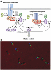
Spatial organization of the R. sphaeroides chemotaxis pathway. (A) The model of signalling in the R. sphaeroides chemotaxis. Attractants are thought to be sensed by the transmembrane chemoreceptors, which regulate activity of CheA2. Signals reflecting the metabolic state of the cell are believed to be sensed by the cytoplasmic receptors, regulating CheA4 and CheA3. CheY6 alone is capable of stopping the flagellar motor, but requires, in addition, either CheY3 or CheY4 to support chemotaxis. CheY6 is mainly phosphorylated by CheA3, but slow phosphotransfer from CheA2 has also been observed (illustrated using thin arrows). The phosphotransfer between CheA2 and CheB2 is reversible (indicated by the bi-sided arrow). (B) Polar and cytoplasmic clusters formed by the two chemosensory complexes. Fluorescence images show localization of the cyan fluorescent protein (CFP) fusion to CheW3 and YFP fusion to CheW4, expressed from endogenous genomic locations.

Comment in

Similar articles

Cited by

References

    1. Adler J, Tso WW (1974) Decision' making in bacteria: chemotactic response of Escherichia coli to conflicting stimuli. Science 184: 1292–1294 - PubMed
    1. Alon U, Surette MG, Barkai N, Leibler S (1999) Robustness in bacterial chemotaxis. Nature 397: 168–171 - PubMed
    1. Amin DN, Hazelbauer GL (2010) The chemoreceptor dimer is the unit of conformational coupling and transmembrane signaling. J Bacteriol 192: 1193–1200 - PMC - PubMed
    1. Armitage JP, Macnab RM (1987) Unidirectional, intermittent rotation of the flagellum of Rhodobacter sphaeroides. J Bacteriol 169: 514–518 - PMC - PubMed
    1. Banno S, Shiomi D, Homma M, Kawagishi I (2004) Targeting of the chemotaxis methylesterase/deamidase CheB to the polar receptor-kinase cluster in an Escherichia coli cell. Mol Microbiol 53: 1051–1063 - PubMed

Publication types

Substances

LinkOut - more resources